Download
1 / 28
280 likes | 299 Views
Learn about Aspergillus fungi, their diverse species, structure, and interaction with the immune system. Discover how cytokine responses, Toll-like receptors, and hyphal growth impact infections.

E N D
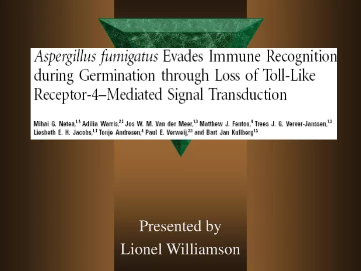
Presented by Lionel Williamson
Aspergillus species • Kingdom: Fungi Phylum: AscomycotaOrder: EurotialesFamily: TrichocomaceaeGenus gi: Aspergillus • Aspergillus is a filamentous, ubiquitous fungus found in nature. It is commonly isolated from soil, plant debris, and indoor air environments.
Types of Aspergillus • The genus Aspergillus includes over 185 species. Around 20 species have so far been reported as causative agents of opportunistic infections in man • A. flavus produces aflotoxins • A. nidulans produces cutaneous aspergillosis • A. oryzae used to make soy sauce, and sake
A. fumigatus • Is the second most common opportunistic fungal infection (Candida) • This species is the most common agent of aspergillosis in both man and animals. It is a thermophilic species (growth at 40°C and beyond) that may be extremely angioinvasive, particularly in the compromised patient
Key words • T helper 1- TH1 Cytokine response that deals with pro inflammatory response • T helper 2- TH2 Cytokine response that deals with anti-inflammatory response • PBMC are macrophages and are involved with the innate immune response • Innate immunity refers to antigen-nonspecific defense mechanisms that a host uses immediately or within several hours after exposure to almost any antigen. This is the immunity one is born with and is the initial response by the body to eliminate microbes and prevent infection.
Cytokines • Proteins produced by white blood cells that act as chemical messengers between cells. They can stimulate or inhibit the growth and activity of various immune cells • Pro-inflammatory response ( TNF, IL-1) • Anti-inflammatory response (IL-10)
Structure of Aspergillus • Conidia are asexual spores that grown on elaborate structures called conidiophores. These are usually stalked, lifting the conidia off the substrate for better dispersal and to avoid microscopic grazing animals. They often produce hundreds or thousands of conidia at a time • Hyphae A hypha (plural hyphae) is a long, branching filament that, with other hyphae, forms the feeding thallus of a fungus called the mycelium. A typical hypha consists of a tubular wall, usually made of chitin, which surrounds, supports, and protects the cells that compose a hypha. For most fungi, a cell within a hyphal filament is separated from other cells by internal cross-walls called septa
Hyphae vs. conidia The University of Adelaide
Life cycle of Aspergillus • A fungus begins its existence as an independent biont as some sort of propagule, most often a spore. From the spore, the fungus grows in a thread-like, branching formation known as a hypha. • The hypha grow and intertwine and form mycelia which become the body of the fungus.
Germinating conidiaRequirement of spermidine for developmental transitions in Aspergillus nidulansYuan Jin 1 , Jin Woo Bok 2 , Doralinda Guzman-de-Peña 3 and Nancy P. Keller
Toll like receptors • Are pattern-recognition receptors believed to play a role in innate immunity • The receptor was first isolated from Drosophila as a gene required for ontogenesis and antifungal resistance • There have been several TLR’s isolated from humans
Types of TLR’s • TLR 2 recognizes peptidoglycan that are associated with gram positive bacteria • TLR 4 recognizes LPS associated with gram negative bacteria • TLR5 recognizes flagella • TLR9 recognizes bacterial DNA
Experiment 1 • Challenged mouse macrophages with conidia or hyphae from A. fumigatus • They found that maximal cytokine release induced by stimulation with 10^7 cfu • Assessed TLR4’s role by using a TLR4-deficient mutant compared to a control • Assessed TLR2’s role by using a TLR2-deficient mutant compared to a control
Experiment 2 • To prove that TLR4 was involved in conidia and not hyphae recognition • Using a NF-kB reporter plasmid that drives CD 25 expression • Used a cell line (3E10) that expressed TLR4 but not TLR2 • Stimulated the cells with LPS (TLR4 antagonist) Pam3cys (TLR2 antagonist) • Then they transfected cDNA for TLR2
Experiment 3 • They wanted to see what roles TLR4 and TLR2 played on the Pro-inflammatory response • Used PBMC’s with antibodies blocking the TLR4 then stimulating with 10^7 cfu • The same experiment was repeated with TLR2
Experiment 4 • Which TLR stimulated IL-10 • Recent data suggests that TLR2 stimulates TH-2 response and TLR4 stimulates a TH-1 response • Role of IL-10 in invasive aspergillosis: increased resistance of IL-10 gene knockout mice to lethal systemic aspergillosis K. V. Clemons, G. Grunig* , R. A. Sobel , L. F. Mirels, D. M. Rennick* & D. A. Stevens
My conclusion • I think that what happens is that Aspergillus conidia gets into the body and meets the macrophages that expresses TLR4. This causes an increase of expression of IL-1 and TNF, this kills the most of the fungus. • The fungus counteracts this by germinating from the conidia to the hyphae forms. TLR4 does not recognize the new form and the hyphae grows. In a normal immune system TLR2 which has 2 functions (pro-inflammatory and anti-inflammatory) with the help of the cell mediated response the body is able to fight off the fungal infection
Immunocompromised • The same things occur but there is a diminished cell mediated response due to the disease process • TLR2 is releasing both pro-inflammatory and an Anti-inflammatory response • This decreases the destruction of the hyphae and they continue to grow. • Where they form mycelia and cause Aspergillosis.
Aspergillosis • - Pulmonary aspergillosis 1- CNS aspergillosis- Sinonasal aspergillosis- Osteomyelitis - Endophthalmitis- Endocarditis- Renal abscesses- Cutaneous • Why do you think pulmonary aspergillosis is the number one form?
This is the air sacs of a hen during an outbreak of aspergillosis The University of Adelaide
Fungal balls made up of hyphae isolated from the lungs The University of Adelaide
Aspergillus fumigatus in lung tissue, note conidial heads forming in an alveolus The University of Adelaide
Aspergilloma found at post-mortem in the lung of a child with leukemia. Note fungus ball occupying cavity The University of Adelaide