Download

1 / 49
1.15k likes | 3.55k Views
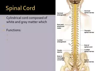
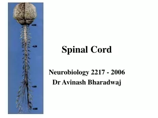
Spinal Cord. Runs through the vertebral canal Extends from foramen magnum to second lumbar vertebra Regions Cervical Thoracic Lumbar Sacral Coccygeal Gives rise to 31 pairs of spinal nerves All are mixed nerves Spinal cord Enlargements Cervical enlargement: supplies upper limbs

E N D
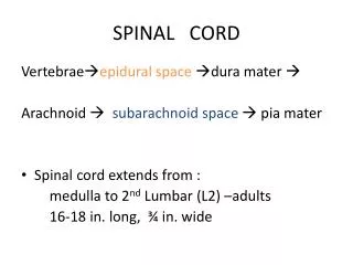
Spinal Cord • Runs through the vertebral canal • Extends from foramen magnum to second lumbar vertebra • Regions • Cervical • Thoracic • Lumbar • Sacral • Coccygeal • Gives rise to 31 pairs of spinal nerves • All are mixed nerves • Spinal cord Enlargements • Cervical enlargement: supplies upper limbs • Lumbo -sacral enlargement: supplies lower limbs • Conus medullaris- tapered inferior end • Ends between L1 and L2
Spinal Segments & Roots Spinal segment C8, T12, L5, S5, Cx1 Anterior (Ventral) Root Posterior (Dorsal) Root Dorsal Root (Spinal) Ganglion Root - Rootlets
Spinal Segments • Importance of the spinal segments
Coverings of Spinal cord • Dura mater: outermost layer; continuous with epineurium of the spinal nerves • Arachnoid mater: thin and web like • Pia mater: bound tightly to surface • Ligamentum Denticulatum • Cordotomy • Forms the filum terminale • anchors spinal cord to coccyx • Spaces • Epidural: external to the dura • Anesthestics injected here • Epidural Anesthesia • Subdural space: serous fluid • Subarachnoid: between pia and arachnoid • Filled with CSF
Lumbar Puncture Lumbar Puncture – lumbar (terminal) cistern
Spinal Cord • White Matter Anterior Funiculus (Anterior White Column) Posterior Funiculus (Posterior White Column) Lateral Funiculus (Lateral White Column) • Gray Matter Anterior Horn ------------ motor Posterior Horn -------------- sensory Lateral Horn ----------------- autonomic (sympathetic) Gray Commissure -------- anterior and posterior
Cord Organization • Principles of Cord Organization 1) Longitudinal Arrangement Fibers (White Matter) ------------ White Column Cell Groups (Gray Matter) ------- Gray Column 2) Transverse Arrangement Afferent & Efferent Fibers Crossing (Commissural and Decussating) Fibers 3) Somatotopical Arrangement
Somatosensory Pathway Posterior column pathway carries sensation of highly localized touch, pressure, vibration. Posterior column pathway includes: • Fasciculus cuneatus tract • Fasciculus gracilus tract - Carries fine touch, pressure, vibration, sterognosis and conscious Proprioceptive sensations.
dorsal cloumn pathway Dorsal Colum tracts
Left spinal cord injury dorsal column pathway • Loss of sense of: • touch • proprioception • vibration • in left leg Dorsal Colum Lesion
Dorsal Colum Lesions Sensory ataxia Patient staggers; cannot perceive position or movement of legs Visual clues help movement Rombergism
Case …. An 85-year-old man is being evaluated for gait difficulties. On examination it is found that joint proprioception is absent in his toes. People with impaired position sense will usually fall if they stand with their feet together and do which of the following? Flex the neck Extend their arms in front of them Flex the knees Turn the head Close their eyes
Clinical Case • A 45 year old woman complained of pain in her right breast and progressive weakness of her right lower limb for a period of two months, she contacted her Family physician, Her Family physician referred her to a neurologist. • The neurologic evaluation revealed weakness in the right lower limb. This was associated with spasticity (increased tone), hyperreflexia (increased deep tendon reflexes) at the knee and ankle, which also demonstrated clonus. • On the right side there was loss of two-point discrimination, touch ,vibratory sense and proprioception at levels below the hip. The left side showed a loss of pain and temperature sensation below dermatome T-7.
Clinical Case Of Spinal Cord cont.. MRI of a patient indicated to have an extramedullary tumor expanding from the dorsal roots at spinal cord levels T-5,6. Based on the symptoms and clinical findings what is your diagnosis ?
The Anterolateral Pathway • Provides sensations of “crude” touch, pressure, pain, and temperature • Ascend within the anterior or lateral spinothalamic tracts:
What is Pain? • “An unpleasant sensory & emotional experience associated with actual or potential tissue damage, or described in terms of such damage” – • Subjective sensation • Pain Perceptions – based on expectations, past experience, anxiety, suggestions • Affective – one’s emotional factors that can affect pain experience • Behavioral – how one expresses or controls pain • Cognitive – one’s beliefs (attitudes) about pain • Physiological response produced by activation of specific types of nerve fibers • Experienced because of nociceptors being sensitive to extreme mechanical, thermal, & chemical energy. • Composed of a variety of discomforts • One of the body’s defense mechanism (warns the brain that tissues may be in jeopardy)
Where Does Pain Come From? • Cutaneous Pain – sharp, bright, burning; can have a fast or slow onset • Deep Somatic Pain – stems from tendons, muscles, joints, periosteum, & b. vessels • Visceral Pain – originates from internal organs; diffused @ 1st & later may be localized (i.e. appendicitis) • Psychogenic Pain – individual feels pain but cause is emotional rather than physical
Left spinal cord injury spinothalamic pathway • Loss of sense of: • Touch • Pain • Warmth/cold • in right leg Anterolateral System (Pain &Temp)
Spinothalamic Tracts • Located lateral and ventral to the ventral horn • Carry impulses concerned with pain and thermal sensations (lateral tract) and also non- discriminative touch and pressure (medial tract) • Fibers of the two tracts are intermingled to some extent • In brain stem, constitute the spinal lemniscus • Fibers are highly somato-topically arranged, with those for the lower limb lying most superficially and those for the upper limb lying deeply
Lateral Spinothalamic Tract • Carries impulses concerned with pain and thermal sensations. • Axons of 1st order neurons terminate in the dorsal horn • Axons of 2nd order neuron (mostly in the nucleus proprius), decussate within one segment of their origin, by passing through the ventral white commissure & terminate on 3rd order neurons in ventral posterior nucleus of the thalamus • Thalamic neurons project to the somatosensory cortex
Anterior Spinothalamic Tract • Carries impulses concerned with non- discriminative touch and pressure • Axons of 1st order neurons enter cord terminate in the dorsal horn • Axons of 2nd order neuron (mostly in the nucleus proprius) may ascend several segments before crossing to opposite side by passing through the ventral white commissure & terminate on 3rd order neurons in ventral posterior nucleus of the thalamus • Thalamic neurons project to the somatosensory cortex
Spino-reticulo-thalamic System • The system represents an additional route by which dull, aching pain is transmitted to a conscious level • Some 2nd order neurons terminate in the reticular formation of the brain stem, mainly within the medulla • Reticulothalamic fibers ascend to intralaminar nuclei of thalamus, which in turn activate the cerebral cortex
Pain Control Theories • Gate Control Theory • Endogenous Opiates Theory • Phantom Pain • Refferd Pain
Gate Control Theory • Melzack & Wall, 1965 • Substantia Gelatinosa (SG) in dorsal horn of spinal cord acts as a ‘gate’ • SG cells of Lamina II act as a inhibitory neurons and inhibit “T” cells of lamina IV • Larger diameter afferent fibers of touch excite both SG and T cells, Therefore afferent signals of pain sensation from T cells is blocked by stimulation of inhibitory SG cells. • Small diameter afferent fibers excite T cells and Inhibit SG cells Therefore Gate is kept
Descending Pain Inhibition • Descending Pain Modulation (Descending Pain Control Mechanism) • Periaqueductal Gray Area (PGA) – release enkephalins • Nucleus Raphe Magnus (NRM) – release serotonin • The release of these neurotransmitters inhibit ascending neurons • Stimulation of the PGA in the midbrain & NRM in the pons & medulla causes analgesia. • Endogenous opioid peptides - endorphins & enkephalins
Referred Pain? • Dermatomal rule • Convergence • Facilitation
Spinocerebellar Tracts • The spinocerebellar system consists of a sequence of only two neurons • Two tracts: Posterior & Anterior • Located near the dorsolateral and ventrolateral surfaces of the cord • Contain axons of the second order neurons • Carry information derived from muscle spindles, Golgi tendon organs and tectile receptors to the cerebellum for the control of posture and coordination of movements
Posterior Spinocerebellar Tracts • Present only above level L3 • The cell bodies of 2nd order neuron lie in Clark’s column • Axons of 2nd order neuron terminate ipsilaterally (uncrossed) in the cerebellar cortex by entering through the inferior cerebellar peduncle
Ventral Spinocerebellar Tracts • The cell bodies of 2nd order neuron lie in base of the dorsal horn of the lumbosacral segments • Axons of 2nd order neuron cross to opposite side, ascend as far as the midbrain, and then make a sharp turn caudally and enter the superior cerebellar peduncle • The fibers cross the midline for a second time within the cerebellum before terminating in the cerebellar cortex • Both spinocerebellar tracts convey sensory information to the same side of the cerebellum
Spinotectal Tract • Ascends in the anterolateral part in close association with spinothalamic system • Primary afferents reach dorsal horn through dorsal roots and terminate on 2nd order neurons • The cell bodies of 2nd order neuron lie in base of the dorsal horn • Axons of 2nd order neuron cross to opposite side, and project to the periaquiductal gray matter and superior colliculus in the midbrain
Spino - olivary Tract • Indirect spinocerebellar pathway (spino-olivo-cerebellar) • Impulses from the spinal cord are relayed to the cerebellum via inferior olivary nucleus • Conveys sensory information to the cerebellum • Fibers arise at all level of the spinal cord
Spinoreticular Tract • Originates in laminae IV-VIII • Contains uncrossed fibers that end in medullary reticular formation & crossed & uncrossed fibers that terminate in pontine reticular formation • Form part of the ascending reticular activating system
Spino-Olivary Tracts • Project to accessory olivary nuclei and cerebellum. • Contribute to movement coordination associated primarily with balance.
Spinotectal Tracts • Project to superior colliculi of midbrain. • Involved in reflexive turning of the head and eyes toward a point of cutaneous stimulation.
Spinoreticular Tracts • Involved in arousing consciousness in the reticular activating system through cutaneous stimulation.
Grey Matter Of Spinal cord White Matter Anterior Funiculus (Anterior White Column) Posterior Funiculus (Posterior White Column) Lateral Funiculus (Lateral White Column) Gray Matter Anterior Horn ------------ motor Posterior Horn -------------- sensory Lateral Horn ----------------- autonomic (sympathetic) Gray Commissure -------- anterior and posterior
Principles of Cord Organization 1) Longitudinal Arrangement Fibers (White Matter) ------------- White Column Cell Groups (Gray Matter) ------- Gray Column 2) Transverse Arrangement Afferent & Efferent Fibers Crossing (Commissural and Decussating) Fibers 3) Somatotopical Arrangement
Principles of Cord Organization Lamina of Rexed Lamina I ---------- posteromarginal nucleus Lamina II ---------- substantia gelatinosa of Rolando Lamina III, IV ----- nucleus proprius Lamina V, VI Lamina VII --------- intermediate gray intermediolateral cell column (ILM) Clarke’s column (Nucleus dorsalis) intermediomedial cell column (IMM) Lamina VIII Lamina IX ---------- anterior horn (motor) cell Lamina X ----------- gray commissure
Alpha Motor Neurons • Motor Unit • Motor End Plate • Phasic • Tonic