Download

1 / 18
190 likes | 661 Views
Learn about biofeedback instruments used in muscle therapy, measuring electrical activity with EMG, electrode placement, and clinical applications. Discover how biofeedback helps with muscle re-education, relaxation, and pain control.

E N D
Biofeedback Chapter 7
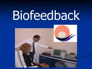
Biofeedback • Electronic or electromechanical instruments that accurately measures, processes, and provides feedback via auditory or visual signals • Used to help patient develop greater voluntary control of either • Neuromuscular relaxation, or • Muscle re-education following injury
Role of Biofeedback • Intrinsic feedback = movement • Kinesthetic, visual, cutaneous, vestibular, and auditory signals • Extrinsic feedback = knowledge • Results presented verbally, mechanically, or electronically to indicate the outcome of some movement performance
Role of Biofeedback • Feedback is ongoing • Occurs before, during, and after any motor or movement task • Feedback from some measuring instrument which provides moment-to-moment information about a biologic function is referred to as biofeedback
Role of Biofeedback • Patient able to make appropriate, small changes in performance which are immediately noted and rewarded • Eventually larger changes, or improvements, in performance can be accomplished • Goal = train patient to perceive changes without the use of a biofeedback unit
Biofeedback Instruments • Measure electromyographic activity (EMG)indicating amount of electrical activity during muscle contraction • Most common type of biofeedback used in athletic training
EMG Biofeedback • Nerve fiber conducts an impulse to the neuromuscular junction where acetylcholine binds to receptor sites on the sarcolemma inducing a depolarization of the muscle fiber • Changes in electrochemical potential difference associated with depolarization can be detected by an electrode placed in close proximity
Measuring Electrical Activity • EMG does not measure muscle contraction directly • Measures electrical activity associated with muscle contraction • Units of measure are microvolts • 1 volt = 1,000,000 µV • EMG readings may be compared only when the same equipment is used for all readings
Measuring Electrical Activity • EMG biofeedback unit receives small amounts of electrical energy generated during muscle contraction (via the electrodes) • Separates or filters electrical energy from other extraneous electrical activity on skin • Amplifies the EMG electrical energy and converts it to some type of information which has meaning to the patient • Meter, auditory signal, light display
EMG Electrodes • Skin surface electrodes • Some electrodes permanently attach to cable wires while others may snap onto the wire • Some units include a set of three electrodes pre-placed on a velcro band which attaches to the skin
EMG Electrodes • Size of electrodes varies • 4 mm diameter for small muscle activity • 12.5 mm diameter for larger muscles • Increasing the size of the electrode will not cause an increase in the amplitude of the signal • Electrodes may be disposable or non-disposable • Require some type of conducting gel
EMG Electrode Placement • Prepare skin by scrubbing with an alcohol-soaked prep pad • Electrodes should be placed as near to the muscle being monitored as possible • Electrodes should be parallel to the direction of the muscle fibers • Spacing of the electrodes is critical to reduce extraneous electrical activity (noise)
Separation and Amplification of EMG Activity • 2 active electrodes • 1 reference electrode • Active electrodes pick up electrical activity from motor units firing in the muscles beneath the electrodes
Converting EMG Activity to Meaningful Information • Biofeedback units generally provide either visual or auditory feedback relative to the quantity of electrical activity • Visual feedback uses lights, bars, or analogue or digital meters • Auditory feedback uses increasing or decreasing tones, buzzing, beeping or clicking
Setting Sensitivity • Sensitivity may be set at… • 1 µV, 10 µV, or 100 µV • A high sensitivity means the biofeedback unit is sensitive enough to detect the smallest amounts of electrical activity • Higher sensitivity levels should be used during relaxation training • Lower sensitivity levels should be used during muscle re-education training
Clinical Applications • Muscle re-education • Isometric contractions sustained for 6-10 sec • Maximize feedback • Tx time = 5-10 min • Relaxation of muscle guarding/Pain control • Concentrate on muscle relaxation • Minimize feedback • Change positions
Indications: • Muscle re-education • Regaining neuromuscular control • Increasing isometric/isotonic strength • Relaxation of muscle spasm/guarding • Pain reduction • Psychological relaxation Contraindications: • Any musculoskeletal condition in which a muscle contraction may exacerbate the condition