Download
1 / 52
600 likes | 971 Views
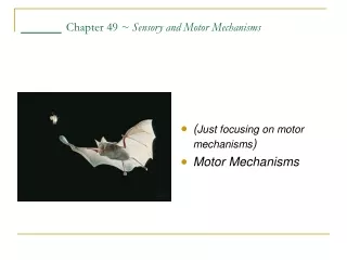
Sensory and Motor Mechanisms. Chapter 49. Sensing and Acting. Bats use sonar to detect their prey Moths c an detect the bat’s sonar and attempt to flee Both of these organisms have complex sensory systems that facilitate their survival. Types of Sensory Receptors.

E N D
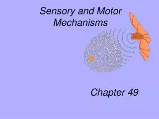
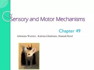
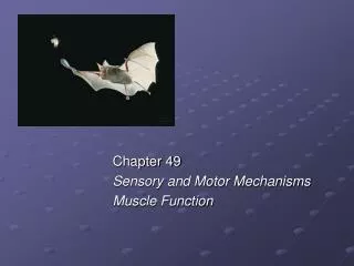
Sensory and Motor Mechanisms Chapter 49
Sensing and Acting • Bats use sonar to detect their prey • Moths can detect the bat’s sonar and attempt to flee • Both of these organisms have complex sensory systems that facilitate their survival
Types of Sensory Receptors • Based on the energy they transduce, sensory receptors fall into five categories • Mechanoreceptors • Chemoreceptors • Electromagnetic receptors • Thermoreceptors • Pain receptors
Mechanoreceptors • Mechanoreceptors sense physical deformation • Caused by stimuli such as pressure, stretch, motion, and sound • The mammalian sense of touch • Relies on mechanoreceptors that are the dendrites of sensory neurons
Chemoreceptors • General receptors that transmit information about the total solute concentration of a solution • Specific receptors that respond to individual kinds of molecules • EX: Taste, Smell
Figure 49.5 Chemoreceptors in an insect: Female silk moth Bombyx mori releasing pheromones; SEM of male Bombyx mori antenna
Electromagnetic Receptors • Electromagnetic receptors detect various forms of electromagnetic energy • Such as visible light, electricity, and magnetism • Some snakes have very sensitive infrared receptors • That detect body heat of prey against a colder background
Figure 49.6 Specialized electromagnetic receptors: Rattle snake with infrared recpters, beluga whale pod
Figure 49.6bx Beluga whale pod • Many mammals appear to use the Earth’s magnetic field lines • To orient themselves as they migrate
Thermoreceptors • Thermoreceptors, which respond to heat or cold • Help regulate body temperature by signaling both surface and body core temperature
Pain Receptors • In humans, pain receptors, also called nociceptors • Are a class of naked dendrites in the epidermis • Respond to excess heat, pressure, or specific classes of chemicals released from damaged or inflamed tissues
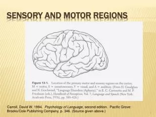
The mechanoreceptors hearing and equilibrium detect settling particles or moving fluid • Hearing and the perception of body equilibrium are related in most animals • Three regions of the human ear • The outer ear • The middle ear • The inner ear
Ear Structure • The outer ear external pinna and the auditory canal • Collects sound and directs it to the tympanic membrane (eardrum)
Middle Ear • Three small bones malleus (hammer), the incus (anvil) and stapes (stirrup) collect vibrations • The eustacian tube equalizes air pressure between the outer and middle ear
Cochlea Stapes Axons ofsensoryneurons Oval window Vestibularcanal Perilymph Apex Base Roundwindow Tympaniccanal Basilar membrane The Cochlea • Snail shaped structure organ of corti
Hearing • Vibrating objects create percussion waves in the air • That cause the tympanic membrane to vibrate • The three bones of the middle ear • Transmit the vibrations to the oval window on the cochlea • These vibrations create pressure waves in the fluid in the cochlea • That travel through the vestibular canal and ultimately strike the round window
The pressure waves in the vestibular canal • Cause the basilar membrane to vibrate up and down causing its hair cells to bend • The bending of the hair cells depolarizes their membranes • Sending action potentials that travel via the auditory nerve to the brain
Senses of Taste and Smell • Are closely related in most animals • The perceptions of gustation (taste) and olfaction (smell) • Are both dependent on chemoreceptors that detect specific chemicals in the environment
Taste in Humans • The receptor cells for taste in humans • Are modified epithelial cells organized into taste buds • Five taste perceptions involve several signal transduction mechanisms • Sweet, sour, salty, bitter, and umami (elicited by glutamate)
Smell in Humans • Olfactory receptor cellsAre neurons that line the upper portion of the nasal cavity • When odorant molecules bind to specific receptors • A signal transduction pathway is triggered, sending action potentials to the brain
Vision in the Animal Kingdom • Two major types of image-forming eyes have evolved in invertebrates • The compound eye and the single-lens eye • Compound eyes are found in insects and crustaceans • And consist of up to several thousand light detectors called ommatidia • Single-lens eyes • Are found in some jellies, polychaetes, spiders, and many molluscs • Work on a camera-like principle
Simplest Eye • The eye cup of planarians provides information about light intensity and direction but does not form images
Vertebrate Eyes • Camera-like they evolved independently and differ from the single-lens eyes of invertebrates • The main parts of the vertebrate eye are • The sclera, which includes the cornea • The choroid, a pigmented layer • The conjunctiva, that covers the outer surface of the sclera • The iris, which regulates the pupil • The retina, which contains photoreceptors • The lens, which focuses light on the retina
Photoreceptors • The human retina contains two types of photoreceptors • Rods are sensitive to light but do not distinguish colors • Cones distinguish colors but are not as sensitive
Figure 49.13 From light reception to receptor potential: A rod cell’s signal-transduction pathway
The effect of light on synapses between rod cells and bipolar cells
The Human Skeleton • Functions in support, protection, & movement • Animal movements result from muscles working against some type of skeleton • The mammalian skeleton is built from more than 200 bones • Some fused together and others connected at joints by ligaments that allow freedom of movement
Muscles contraction Move Skeletal Parts • The action of a muscle always to contract • Skeletal muscles are attached to the skeleton in antagonistic pairs • With each member of the pair working against each other
Vertebrate Skeletal Muscle • Is characterized by a hierarchy of smaller and smaller units • A skeletal muscle consists of a bundle of long fibers • Running parallel to the length of the muscle • A muscle fiber • Is itself a bundle of smaller myofibrils arranged longitudinally • Skeletal muscle is also called striated muscle • Because the regular arrangement of the myofilaments creates a pattern of light and dark bands
The myofibrils are composed to two kinds of myofilaments • Thin filaments, consisting of two strands of actin and one strand of regulatory protein • Thick filaments, staggered arrays of myosin molecules • Each repeating unit is a sarcomere • Bordered by Z lines • The areas that contain the myofilments • Are the I band, A band, and H zone
The sliding-filament model of muscle contraction • The filaments slide past each other longitudinally, producing more overlap between the thin and thick filaments • As a result of this sliding • The I band and the H zone shrink • The sliding of filaments is based on • The interaction between the actin and myosin molecules of the thick and thin filaments • The “head” of a myosin molecule binds to an actin filament • Forming a cross-bridge and pulling the thin filament toward the center of the sarcomere
One hypothesis for how myosin-actin interactions generate the force for muscle contraction
Hypothetical mechanism for the control of muscle contraction
Types of Skeletons • The three main functions of a skeleton are • Support, protection, and movement • The three main types of skeletons are • Hydrostatic skeletons, exoskeletons, and endoskeletons
Hydrostatic Skeletons • A hydrostatic skeleton • Consists of fluid held under pressure in a closed body compartment • This is the main type of skeleton • In most cnidarians, flatworms, nematodes, and annelids • Annelids use their hydrostatic skeleton for peristalsis • A type of movement on land produced by rhythmic waves of muscle contractions