Download
1 / 58
680 likes | 1.35k Views
Clinical anatomy & physiology of the ear YANG Jun, MD, Ph.D. 09/18/09. Otology & neurotology. Surgical management on hearing loss

E N D
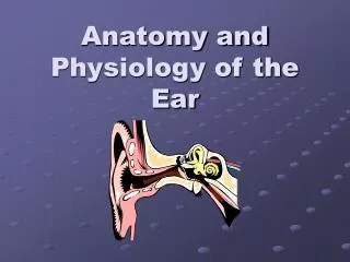
Clinical anatomy & physiology of the ear YANG Jun, MD, Ph.D. 09/18/09
Otology & neurotology • Surgical management on hearing loss • Conductive hearing loss: tympanoplasty, ossicular chain reconstruction, stapes surgery • Sensorineural hearing loss : implantable hearing-aids, cochlear implatation • Tumor in the lateral skull base,such as acoustic neuroma • Facial nerve: facial paralysis, facial spasm • Surgical management on vertigo • Trigeminal neuralgia • Repaire of CSF leakage
Temporal bone • Location : lateral skull • Neighbour : parietal bone, sphenoid bone, occipital bone • Composition: squamous part, tympanic part, pars mastoidea, petrosal part
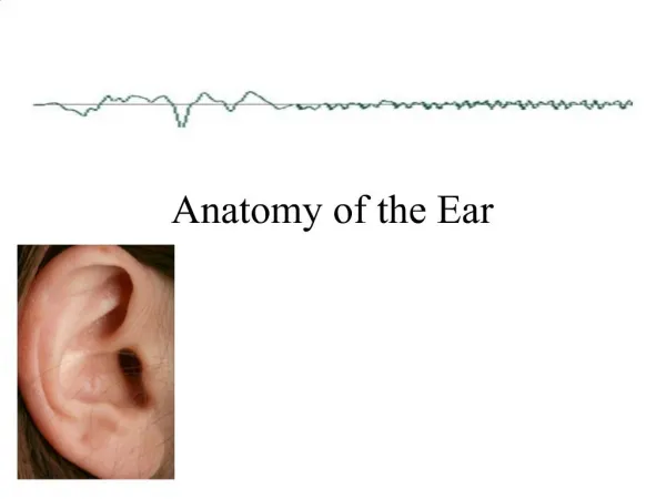
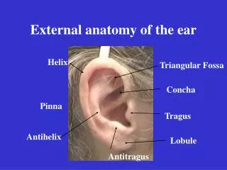
Anatomy of the external ear • auricle • anterior notch of ear-an incision can be made • less subcutaneous tissue • difficult absorption of hematoma • prone to cold injury
Anatomy of the external ear • external auditory canal • 2.5-3.5cm • outer1/3:cartilage • inner2/3:bone • Stenosis: juncture of bone and cartilage, bony part (0.5cm from the tympanic anulus)
Anatomy of the middle ear • Tympanic cavity • Eustachian tube • Tympanic sinus • Mastoid cavity
Tympanic cavity • Attic, mesotympanum, hypotympanum • Six walls: interior, exterior, anterior, posterior, superior, inferior
镫骨底板 面神经管凸 外半规管凸 匙突 鼓窦入口 大脑颞叶 锥隆起 鼓索神经孔 鼓岬 面神经 砧骨 锤骨 鼓膜张肌半管 鼓膜张肌附着处 咽鼓管鼓口 鼓索神经 鼓膜 颈内动脉 蜗窗小窝 颈静脉球 Tympanic cavity
Exterior wall-tympanic membrane • Tympanic membrane • Semi-transparent film, 1cm2, 1mm • Upper is pars flaccid, lower is pars tensa • Three layer construction: epithelial lamina, fibrous lamina, mucous layer
Interior wall • Namelyexterior wall of the inner ear • Center-promontorium tympani • Post-superior : vestibular window-vestibule • Post-inferior : cochlear window-scala tympani • horizontal part of facial nerve canal • prominence of lateral semicircular canal • cochleariform process
Anterior wall • Namely carotid wall • Inferior part is separated with the carotid artery • Two openings at the superior part: semicanal for tensor tympani (upper), semicanal for auditory tube (lower)
Posterior wall • Minipore at the posterior wall- aditus ad antrum tympanicum • incudal fossa- juncture of horizontal part and perpendicular part • pyramidal eminence-about at height of vestibular window • facial recess-posterior tympanotomy
Superior wall • Namely tegmen tympani • Be separated with the temporal lobe of the cerebrum in the middle fossa • The petrosquamous fissure in infant is not closed-one of the route by which infection from the middle ear could get into
Inferior wall • Namely jugular wall • Be separated with the jugular bulb • blue drum
Content in the tympanic cavity • ossicles(smallest bone in the human body): malleus, incus, stapes- ossicular chain • ligamenta ossiculorum auditus: ligament of the malleus, incus and stapes • muscle in the tympanic cavity: tensor tympani muscle, stapedial muscle • chorda tympani nerve
Eustachian tube • Passageway between tympanic cavity and nasopharynx, outer 1/3-bony part, inner 2/3- cartilaginous part. Isthmic portion-junction of bony part and cartilaginous part. • The opening at the nasopharynx is open when muscle contraction in order to adjust air pressure in the tympanic cavity. • Infection is prone to enter the tympanic cavity because of Horizontal, short and wide Eustachian tube in child.
Tympanic sinus and mastoid cavity • Tympanic sinus: pneumatic space and passage between the attic and mastoid cavity • Mastoid cavity: cells in the temporal bone-pneumatic type, diploetic type, constrictive type and mixed type
Anatomy of the inner ear • Also labyrinth, containing apparatus responsible for hearing and balance • The inner ear is divided into bony labyrinth and membranous labyrinth • Perilymph is full of the space between bony labyrinth and membranous labyrinth, endolymph is full of the membranous labyrinth
bony labyrinth • Compact bone • Vestibule, semicircular canal, cochlea
Vestibule • Between the cochlea and the semicircular canal • Five openings from three bony semicircular canals • saccular recess, utricular recess • Exterior wall- vestibular window: sealed by footplate of the stapes
Bony semicircular canals • Three curved bony ducts that form right angle mutually- lateral, superior and posterior semicircular canal • A common crus is formed by the superior and posterior semicircular canal, therefore, five openings from three semicircular canals enter the vestibule
Membranous labyrinth • Composed of membranous duct and membranous sac • fixation at bony labyrinth by fiber bundle • dividing into utricle, saccule, membranous semicircular canal and membranous cochlea (scala media) • cross-connection each other
Membranous labyrinth • Utricle • Utricular recess • Macula utriculi-sense of balance • Five openings in the posterior wall connect with three semicircular canals • Connection with the utriculosaccular duct and endolymphatic duct in the anterior wall. Vestibular aqueduct. Endolymphytic sac (within dura behind the petrosal part of the temporal bone)
Membranous labyrinth • Saccule • Saccular recess • Macula sacculi-sense of balance • Connection with utriculosaccular duct and endolymphatic duct
Membranous labyrinth • Membranous semicircular canal Connection with the utricle
Membranous labyrinth • Membranous cochlea (scala media) • Between the osseous spiral lamina and the lateral wall of the osseous cochlear canal, also between scala vestibuli and scala tympani, containing endolymph • Basilar membrane: from free edge of the osseous spiral lamina • Organ of Corti : hearing receptor composed of outer hair cells and inner hair cells
Physiology of the ear • Hearing • Balance
Route of sound conducting • Air conduction Sound wave auricle external auditory canal vestibular window perilymph/endolymph organ of Corti auditory nerve nucleus auditory cortex
Route of sound conducting • Bone conduction • Sound wave makes the perilymph vibrate through skull route, then stimulates the organ of Corti by which hearing generate. • Translatory mode of bone conduction • Compressional mode of bone conduction
Physiological functions of the external ear • Gathering sound • Discriminating direction • Resonance • Protection • Sound wave pressurizing
Physiological functions of the middle ear • Transformation and gain • Structure for sound transmission and transformation: tympanic membrane and ossicular chain
Physiological functions of the tympanic membrane • Valid area of vibration : 55 mm2 • Area of the footplate: 3.2 mm2 17times
Function Middle ear—amplification from area ratio • Pressure = Force/area • Area of tympanic membrane ~17 > area stapes • Gain of area ratio ~24 dB
Physiological functions of the ossicular chain • Lever manubrium of malleus long crus of incus 1.3:1 • 1.3×17=22.1 27dB
Function of Middle ear—pressure amplification-ossicles • Energy loss at air-fluid interface-99.9% loss (-30 dB) • Malleus longer than incus-amplify pressure ~1.7X (+2 dB)
Physiological functions of muscles in the tympanic cavity • stapedial muscle: decreasing pressure of perilymph
Physiological functions of muscles in the Eustachian tube • Keeping balance of pressure in the middle ear • Drainage • Prevention of retrograde infection • Noise abatement