Download
1 / 56
590 likes | 1.02k Views
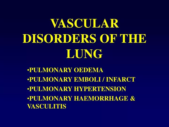
VASCULAR DISORDERS OF THE LUNG. PULMONARY OEDEMA PULMONARY EMBOLI / INFARCT PULMONARY HYPERTENSION PULMONARY HAEMORRHAGE & VASCULITIS. PULMONARY EMBOLI / INFARCT. PE – most common preventable cause of death in hospitalized patients

E N D
VASCULAR DISORDERS OF THE LUNG PULMONARY OEDEMA PULMONARY EMBOLI / INFARCT PULMONARY HYPERTENSION PULMONARY HAEMORRHAGE & VASCULITIS
PULMONARY EMBOLI / INFARCT • PE – most common preventable cause of death in hospitalized patients • Embolus: A detached intravascular solid, liquid or gaseous mass that is carried by the blood to a site distant from its point of origin • 99% of all emboli are thromboemboli • Rarer – bone, marrow, atheroma, fat, tumour, FBs [ cotton, cardiac catheter, talc (ivdu)], parasites, amniotic fluid • 95% PE arise in thrombi in large deep veins of LL • What happens depends on size:occlude main PA, lodge at bifurcation = saddle embolus, shower of smaller emboli may travel distally, passage thru ASD/VSD = “paradoxical embolus”
PULMONARY EMBOLI / INFARCT • Most PEs are small and silent • Respiratory & haemodynamic compromise • If > 60% of total pulmonary vasculature obstructed - sudden death, acute RHF, EMD • Middle size arteries – haemorrhage • Obstruction of smaller Pul. Aa branches (end arteries) - infarction • PE leading to infarction uncommon in young – but is seen where circulation already inadequate – heart & lung disease • Chronically - Pulmonary hypertension, R heart strain
PULMONARY EMBOLI / INFARCT • Acute thromboemboli - blood, fibrin, platelets, neutrophils arranged in alternating linear zones – Lines of Zahn • After 2-3 days – organization, ingrowth of fibroblasts , capillaries from vessel wall • Thrombus is replaced by fibrosis and small vascular spaces – recanalization
PULMONARY EMBOLI / INFARCT • ¾ infarcts affect the lower lobes • Not usually excised – unless clinically unsuspected e.g. unresolving infiltrate or nodular opacity • Classically a wedge shape with base on pleural surface • Central bland necrosis with ghosts of lung architecture, haemorrhage, active fibroblasts at edge, squamous metaplasia, reactive atypia • Eventually a fibrous scar • Consider causes other than simple TE
PULMONARY HYPERTENSION • Mean pulmonary artery pressure >25mmHg at rest, > 30mmHg during exercise • Elevated pressure is related to high pulmonary vascular resistance due to obstruction of small arteries • 3 factors contribute to small pulmonary artery obstruction: vasoconstriction, cellular proliferation & fibrosis and thrombosis • Reclassification at Venice 2003
PULMONARY HYPERTENSIONClinical Classification • Pulmonary arterial hypertension • Pulmonary hypertension with left heart disease • Pulmonary hypertension with lung diseases / hypoxaemia • Pulmonary hypertension due to chronic TE disease
PRIMARY PULMONARY HYPERTENSION • Primary plexiform arteriopathy • Young women (20-40 years) • Dyspnoea & fatigue, some chest pain • Progression to resp distress, RVH and cor pulmonale • Rx – vasodilators, anticoagulants, prostacyclins
SECONDARY PULMONARY HYPERTENSION • Cardiac disease • Obstruction of main Pulmonary Veins • Chronic embolic disease • Lung disease • Alveolar hypoxia • Liver disease , portal hypertension • HIV infection • Ingestants / inhalants • Collagen vascular disease
PULMONARY HYPERTENSIONClinical Classification • Pulmonary arterial hypertension • Pulmonary hypertension with left heart disease • Pulmonary hypertension with lung diseases / hypoxaemia • Pulmonary hypertension due to chronic TE disease
PULMONARY HYPERTENSION Heath and Edwards Grades I medial hypertrophy II intimal proliferation – mild III intimal fibrosis – moderate IV plexiform or dilatation lesions, necrotizing arteritis – severe
PULMONARY HYPERTENSIONPathological classification – Venice 2003 • Pulmonary arteriopathy Medial hypertrophy, isolated or with intimal proliferation, concentric laminar, eccentric, adventitial fibrosis, plexifiorm and / or dilatation lesions, arteritis • Pulmonary occlusive venopathy • Pulmonary Microvasculopathy
PULMONARY HAEMORRHAGE&VASCULITIS ALVEOLAR HAEMORRHAGE SYNDROMES • Goodpasture’s syndrome = Antibasement membrane disease • Idiopathic pulmonary haemosiderosis (IPH) • Wegener’s granulomatosis (WG) • CVD esp. acute Lupus • Drugs, inhalants • Idiopathic RPGN SECONDARY ALVEOLAR HAEMORRHAGE LOCALIZED HAEMORRHAGE
WEGENER’S GRANULOMATOSIS (WG) TRIAD • Granulomatous inflammmation of URT & LRT • Generalized vasculitis • Glomerulonephritis • LUNG most frequently affected • Middle aged adults – but wide age range • Fever, malaise, wt loss, cough, chest pain, hemoptysis, renal failure, anaemia, sinusitis • Radiology – multiple lung masses resembling mets or cavitating abscesses • Serology – Antineutrophil cytoplasmic antibodies (ANCA)
WEGENER’S Multifocal ischaemic necrosis has resulted in numerous cavitating lesions scattered throughout upper and lower lobes of left lung. Dark haemorrhagic lung parenchyma between the cavities to the upper right
ANCA - fluorescent microscopy C-ANCA, diffuse granular cytoplasmic staining pattern in WG P-ANCA, perinuclear staining pattern in microscopic polyangiitis
WG in the LUNG • Necrotizing granulomatous inflammation • Necrotizing vasculitis • Large geographic areas of necrosis (dirty / basophilic / blue under the microscope) • Microabscesses – neutrophils • MNGCs • Ddx: mycobacterial & fungal infection