Download
1 / 15
170 likes | 372 Views
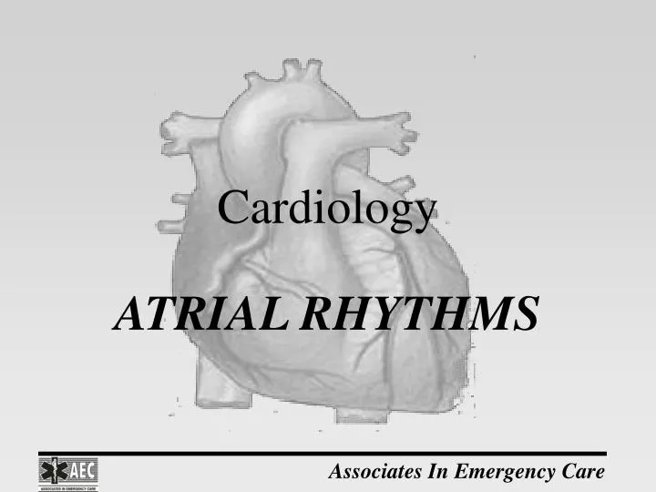
Cardiology. ATRIAL RHYTHMS. Premature Atrial Complexes. Rate: Dependent on underlying rhythm. Rhythm: The regularity of the underlying rhythm is disrupted by the premature complexes.

E N D
Cardiology ATRIAL RHYTHMS
Premature Atrial Complexes • Rate: Dependent on underlying rhythm. • Rhythm: The regularity of the underlying rhythm is disrupted by the premature complexes. • P Wave: The configuration of the premature complex will appear different from the dominant rhythm. The P wave may be hidden in the preceding T wave. • PRI: May be normal or prolonged and differs from the interval of the underlying. • QRS: < .12 seconds
Premature Atrial Complexes • PACs may conduct to the ventricles or be blocked. • This is dependent on where the premature P wave strikes the refractory period of the heart. • When the P wave occurs in the relative refractory period the impulse is conducted. • When the P wave occurs in the absolute refractory period the impulse is blocked and not followed by a QRS complex.
Atrial Tachycardia (SVT) • Rate: 150-250 • Rhythm: Regular • P Wave: Differs in configuration from the Sinus P Wave. May be hidden in the preceding T wave because of the rapid rate. Pacemaker site is a focus in the atrium. This focus overrides the SA Node because its rate is faster than the SA Node. • PRI: Between 0.12 – 0.20 seconds. May not be measurable if the P wave is buried in the preceding T wave. • QRS:< .12 seconds
Atrial Tachycardia (SVT) • This type of tachycardia occurs in two different parameters: 1) Nonparoxysmal Atrial Tachycardia (SVT): this occurs when the tachycardia is the initial presenting rhythm, when the patient is connected to the ECG monitor. 2) Paroxysmal Atrial Tachycardia (SVT): this occurs when you see the change from an initial rhythm to the atrial tachycardia (SVT).
Atrial Tachycardia (SVT) • Atrial Tachycardia is one of the rhythms that we categorize as Supraventricular Tachycardia (SVT). • You may not be able to distinguish where the rhythm is being generated from due to the fast rate. • When the rhythm is regular with normal QRS complexes, we know that the rhythm is being generated from somewhere above the ventricles. • Be definition this is SVT.
Atrial Flutter • Rate: Atrial: 250-350 Ventricular: Depends on the conduction ratio between the atria and ventricles. • Rhythm: Regular or Irregular • P Waves: Atrial deflections are known as F Waves an have a sawtooth appearance. Once ectopic pacemaker site. • PRI: Not measurable (no P waves) • QRS: < .12 seconds
Atrial Fibrillation • Rate: Atrial: 350-600 (chaotic baseline) Ventricular:dependent on the number of conducted impulses. Less than 60 is considered a slow ventricular response. Greater than 100 is considered a rapid ventricular response. 60-100 is considered a controlled ventricular response. NOTE: With atrial flutter and fib it is the ventricular rate that we are treating, not the atrial rate. If bradycardic then treat as bradycardia; if tachycardic then treat as tachycardia. If 60-100 the patient is probably stable and requires no treatment.
Atrial Fibrillation • Rhythm: Irregularly irregular. There are numerous ectopic sites within the atria that are acting as the pacemaker. Independently they are unable to conduct an impulse to the ventricles for the QRS complex. When they collectively fire they are able to conduct an impulse to the ventricles to form the QRS complex. • P Waves: Known as f waves. • PRI: Not measurable (no P waves) • QRS: < .12 seconds
Wandering Atrial Pacemaker • Rate: Varies on shifting pacing sites. • Rhythm: Slightly irregular. • P Waves: Configuration varies according to the dominant pacemaker at the time. Pacemaker site shifts back and forth between SA node and AV junction. • PRI: Depends on the dominant pacemaker. Can be normal or less than 0.12 seconds. • QRS: < .12 seconds