Download

1 / 59
610 likes | 657 Views
This comprehensive guide covers the definition, epidemiology, etiology, clinical diagnosis, treatment, and prognosis of Acute Myeloid Leukemia (AML), including key aspects such as cytogenetics and molecular genetics. It provides insights into the classification, immunophenotyping, and prognostic factors of AML, with a focus on important mutations and therapeutic approaches. The text is based on the latest research and findings in the field by Assistant Lecturer Ahmed Refaat Abd Elzaher from South Egypt Cancer Institute, Assuit University.

E N D
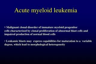
Acute Myeloid Leukemia By Ahmed Refaat Abd Elzaher Assistant lecturer of Medical Oncology South Egypt Cancer Institute Assuit University
Definition & epidemiology • Heterogeneous clonal stemcell malignancy • Immature hematopoietic cells proliferateand accumulate in BM, PB & other tissues featuresof BM failure. • PB and/or BM blasts > 20 % of nucleated cell (NC) count • If the blasts in the BM are > 5% & < 20%MDS • Recently, certain CyG abnormalities classified as AML irrespective of blast count : t(8;21), inv(16)and t(15;17) Roboz G & Sung L. ASH-SAP 2010; PP 475
Acute myeloid leukemia • Incidence – 2.7 per 100,000 • median age of presentation : 65 yo • AML comprises 90% of all acute leukemiasin adults • More prevalent: • Males • European • Hispanic (promyelocytic leukemia (AML M3)
AML - Etiology • Primary AML • Increased incidence • Genetic fragility • Faconi anemia • Down syndrome • tobacco use? • herbicides?, pesticides? • benzene exposure • Secondary AML • XRT • Topoisomerase II inhibitors (e.g etopisode), alkylating agents • MDS • other cell proliferation disorders • CML, polycythemia vera, primary thrombocytosis, PNH
ACUTE LEUKEMIA Clinical Diagnosis Morphological Immunophenotyping Cytogenetics Molecular genetics
Clinical presentation • Sign or symptoms related to pancytopenia: • WBCinfection. • Hb anemia. • platelets bleeding • Approximately 25% of AML patients present with white blood cell (WBC) counts > 50,000/mm³ • Monocytic variants (M4 or M5) commonly display infiltration of gingivae, skin, soft tissues or meninges • DIC is a common presentation in AML M3 • M2 :Chloroma:-presents as a mass lesion ‘tumor of leukemic cells’
Leukostasis • Leukostasis – predominantly in those with WBC counts > 100,000 (10% of patients); can also be seen in patients with WBC > 50,000 • Most common in those with M4 or M5 leukemia • Common symptoms • Pulmonary: dyspnea, chest pain • CNS: headaches, altered mentation, CN palsies, ocular symptoms • Priapism • Myocardial Infarction
Gingival Infiltration in Monocytic AML (M4) After tt !
Diagnosis • Previously >30% blasts on BM aspirate (per FAB criteria)¹ • Recently changed to > 20% blasts in the peripheral blood or BM (per WHO criteria)² • Patients with certain cytogenic abnormalities are considered to have AML regardless of blast percentage² • t(8;21) (q22;q22), inversion (16) (p13q22) • t(16;16) (p13;q22), and t(15;17) (q22;q12) 1- Bennett et al, Br J Haematol 1976 2-Vardiman et al, blood 2002
FAB Classification of AML (1976) 20% 30% 2-4%
AML (WHO 2008) Cytogenetics & Moleculars FAB ! Excluding APL (M3) EXTREMELY IMPORTANT More Easily…
AML: ClassificationWorld Health Organization (WHO) WHO classification (2008) Poor prognosis… BAD cytogenetics AML with MDS Therapy-related AML …. (eg alklating agents, topoisom. II inhib., RT…) (FAB excluding APL)
“Therapy-related AML: AML MM
Cytochemistry ______________________ALL____________AML__________ • Myeloperoxidase - + • Suddan black - + • Non-specific esterase - + in M4, M5 • Periodic acid Schiff (PAS) + in B-ALL + in M6 • Acid phosphatase + in T-ALL + in M6 Bennett et al, Br J Haematol 1976
Immunophenotype Based on blast surface antigen expression CD34 Stem cell marker CD117, CD33, CD13, MPO Myeloid markers CD14, CD64 Monocytic marker Glycophorin A Erythroid marker CD41, CD61 Megakaryocytic markers
AML: Prognostic FactorsFrequency of Cytogenetic Risk Groups Cytogenetic risk groups Roboz G & Sung L. ASH-SAP 2010; 475-487 Jabbour E et al. Mayo Clin Proc. 2006; 81:247 (CBF) Normal / t(9;11) So, practically… (Trisomy 8) Others: Complex (≥3 abnorm.), monosomalkaryotype 5 or 7 (MK 5 / 7 seems to be of the worst !) t(9;22), t(6;11), abn(17).
AML in pts less than 60 years of age: Survival, by karyotype, of de novo and therapy-related (t-AML) Grimwade D & Hills RK. Hematology ASH Educ Program 2009; 385–395. Rowe JM et al. Blood 2010;116:3147
Molecular genetics: The field of biology that studies the structure and function ofgenes at a molecular(atomic)level Moleculars Cytogenetics (CyGs)
“The most important prognostic indicators in AML are age, cytogenetics, and molecular genetics….”
AML: Molecular Mutations and Prognosis Molecular mutations and prognosis (Normal & good cytogenetics… BAD is bad !!!) (Intermediate risk) GOOD GOOD The WORST 20-30% of t(8;21) & 30-40% of inv16 (BAD) & inv16
OS for AML patients with NC according to CEBPA mutation status (similar results in two studies) Preudhomme C et al. Blood 2002; 100:2717 Frohling S et al. JCO 2004; 22:624
cytogenetic markersrevolutionized therapeutic decisions favorable, intermediate & unfavorable. However, patients tend torespond differentlyto a specifictherapeutic approach within the same cytogenetic risk group`. Molecular markers of AML. Remember… • Both the NCCN and the ELN guidelines recommend: molecular genetics (mainly FLT3, NPM1, C-Kit and CEBPA) to further categorizecytogenetically normal patients…. More individualized decision making !
DNMT3A Mutations Common in Intermediate-Risk AML Mutations in the DNA methyltransferase gene, DNMT3A, are also associated with poor prognosis and are highly recurrent in patients with intermediate-risk AML. Presence of any DNMT3A mutation, either alone or in combination with the FLT3 internal tandem duplication (ITD) mutation, is associated with significantly shorter overall survival Ley TJ, et al. ASH 2010. Abstract 99.
Molecular markers of AML Isocitrate dehydrogenase (IDH ) :IDH1 and IDH2 mutations occur in 25–30% of patients with CN-AML, and in general predict for worse outcome in certain molecular (NPM1 wild-type) and clinical (older age) subsets of patients
Evaluation ● Cytogenetics are critical. ● Molecular diagnostics mandatory for all patients including the elderly: at least FLT3-ITD, NPM1,CEBPA; also consider C-KIT in patients with core-binding factor AML. ● Post-remission monitoring of minimal residual disease using cytogenetics, PCR, and multicolor flow cytometry. ● Send HLA typing of patient and siblings at time of initial diagnosis.
Assessment of response to treatment CALGB Criteria Detection of minimal residual disease (MRD) CR PR MRD- MRD+
Criteria of response to treatment (CALGB) Definition of CR Clinical Peripheral blood Bone marrow • P.S. = 100% • No organomegaly • TLC= 2-10 X 109/L • ANC > 1000 • No blasts • HB > 11 (females) • > 12 (males) • PLT > 100 000/m3 • Normocelluar • Blasts <5% The presence of Auer rods is not allowed in CR marrow NCCN Guidelines Version 2.2011
AML: Initial Treatment …previously !! Essentially, routine practice nowadays for all AML pts !
Treatment of AML 1) Supportive treatment (for all pts) 2) Induction therapy 3) Post-remission (consolidation) therapy 4)Treatment of Relapsed AML • 5) Special considerations • Elderly AML • Concept of targeted therapy in AML
Supportive treatment • Hydration and allopurinol to prevent tumor lysis syndrome… esp initial cyto-reduction with HU (if TLC) • Availability of “full” neutropenic precautions • Insertion of a central line to deliver chemotherapy . • Blood and platelets transfusion • Oral care and nutritional advice • Antimicrobials (prophylactic / therapeutic) • Sperm banking • Hormonal therapy to control menses during time of thrombocytopenia.
To begin with, till this very moment…. It is still the: “3 + 7” regimen (from the 1970s …..!!) Anthracycline X 3d (usually Daunorubicin) AraC (Cytarabine) X 7d Although AML…heterogeneous Same tt approach (except APL)… !
Induction Treatment Of AML Standard DA (3+7) BUT … Initially • Dauno 45mg/m2 D 1-3 I.V • AraC100 / 200 mg/m2 D 1-7 C.I No Difference in OS
SO… (Fernandez HF et al. NEJM 2009; 361:1249)
Which anthracycline ? Idarubicin (10 or 12mg/m2 X 3d) OR Mitoxantrone 12mg/m2 X 3d NO advantage over the higher doses of Dauno (i.e 60 or 90)
Ok, 3+7…. then what ?!
Still with remission induction Day 14 BM Blasts <5% wait till CBC recovery Blasts >5% & < 50% of initial blasts 2nd 3+7 (if tolerable & NO active infection) Blasts > 50% of initial blasts Salvage (eg HAM / FLAG-M) Upon CBC recovery BM (Nadir)
Treatment of AML 1) Supportive treatment (for all pts) 2) Induction therapy 3) Post-remission (consolidation) therapy 4) Treatment of Relapsed AML • 5) Special considerations • Elderly AML • Concept of targeted therapy in AML
Consolidation therapy: “EXTREMELY” crucial….. AML: Treatment Overview • Rational: eradicate residual Leukemic clone Thus, traditionally…
Consolidation after CR(exclusively… cytogenetics) BUT, currently…. Good risk HiDAC x 3-4 Allo-HSCT OR HiDAC AML (1st CR) Intermed. risk Allo-HSCT OR clinical trial Poor risk • Two meta analyses superiority of Allo in these risk groups: • Cornelissen J et al. Blood 2007; 109:3658 • Koreth A. et al. JAMA 2009; 301:2349
Current facts The new era of molecular incorporation…. therapeutic map is changing (Few grey zones...!)
Favorable Intermediate Unfavorable No role for HSCT,EXCEPT: 45% of AML, mostly cytogenetically normal (CN) Allo-HSCT for all Those with a mutation inc-KIT (eg with CBF t 8;21) or FLT3 Allo-HSCT for allEXCEPTmutant NPM1 but FLT3-ITD -veORCEBPA+ve
Post-remission therapy for adults with newly diagnosed AML (except APL) Induction by (3+7) irrespective of risk group CR Favorable risk Intermediate/unfavorable risk HD ARA-C containing regimen X 3 cycles Donor present No donor HD ARA-C containing regimen X 3 Relapse Allo BMT in CR1 as soon as possible 2nd induction ABMT in CR1 Donor present No donor Relapse Allo BMT in CR2 ABMT in CR2 Palliative therapy
Treatment of special forms of AML AML M5 High Risk Of CNSDisease After reaching CR by induction chemotherapy, give triple intrathecal prophylaxis: • MTX 15 mg • ARA-c 40 mg • Dexamethazone 4 mg Every 8 weeks for a total of 6 injections.