Download
1 / 20
200 likes | 223 Views
Explore the divisions and structures of the brain including the telencephalon, diencephalon, mesencephalon, and rhombencephalon. Learn about the functions of thalamus, hypothalamus, pons, medulla, spinal cord, and more.

E N D
Chapter 2b CNS Gross Anatomy • Chris Rorden University of South Carolina Norman J. Arnold School of Public Health Department of Communication Sciences and Disorders University of South Carolina
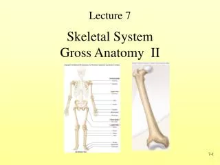
Brain Sub-Divisions Telencephalon – Cortex, Limbic system, basal ganglia, olfactory bulb Diencephalon Thalamus, Hypothalamus Mesencephalon Tegmentum, Red Nucleus, Colliculi Rhombencephalon Pons, Cerebellum, Medulla Spinal Cord
Diencephalon • Diencephalon = thalamus, hypothalamus, epithalamus, prethalamus or subthalamus and pretectum • Thalamus • Oval Mass of Neurons in the Center of the Brain • Relays Sensorimotor Information to the Cortex
Thalamus • Poor contrast on MRI scans – hard to see.
Thalamic Injury • T2-weighted MRI • Bilateral injury to parts of thalamus
Diencephalon • Hypothalamus • Body Heat, Water intake, Hormone Production, Emotional Expression, Food Consumption and Reproduction
Midbrain, Hindbrain • Pons • Medulla Oblongata • Functions: Automatic Control Systems
Ventral Brainstem • Mamillary Bodies • Medial Sulcus • Ventral Median Fissure • Pyramidal Tract • Pyramidal Decussation • Where signals cross • Ventral lateral sulcus
Midbrain Dorsal Structures • Superior and Inferior Colliculus • The ‘little hills’. • Superior colliculi – help initiate eye movements, integrate visual, auditory and tactile information • Inferior colliculi- audition • Aka Corpora Quadrigemina (four seeds) Dorsal View of Brainstem Pineal Body Brachium Superior Colliculus Inferior Colliculus Colliculus Superior Inferior Trochlear Nerve
Colliculi • Superior colliculi: eye movement control. Also maps touch and hearing onto vision • Inferior colliculi: pathway for auditory processing Sagittal View
Internal Structures of the Midbrain • Tegmentum • Cerebral Aqueduct • Red Nucleus • Cranial nerve nuclei • Reticular Formation • Central Gray Area Transverse Section of Rostral Midbrain Tectum Tegmentum Basis Pedunculi
Internal structures of the pons • Tegmentum • Ascending and Descending Fibers • Medial Lemniscus • Basilar Pons • Cortical Descending Tracts • Pontine Nuclei • Pontocerebellar Fibers Transverse Section of Mid Pons Teg- mentum Basilar
Medulla Oblongata • Connects to Pontine Protrusion • Caudal Portion Contains Pyramidal Decussation • Where motor tracts cross over for control of contralateral side of the body Tegmentum Pyramid
Landmarks of Medulla • Ventral • Ventrolateral Sulcus • Pyramidal Tract • Lateral Corticospinal Motor Tract • Dorsal • Dorsal Median Sulcus • Dorsolateral Sulcus • Olivary Nucleus
Internal Medulla • Dorsal Tegmentum • Ventral Pyramidal Tract • Reticular Formation • Cardiac Center • Vasomotor Center • Respiratory Center
Cerebellum • Modifies Cortical Activity • Distinctive Appearance • Gyri called Folia • Two Hemispheres • Three Lobes • Anterior • Posterior • Flocculonodular
Cerebellum Posterior lobe • Rostral view • (bird’s eye) Anterior lobe
Cerebellum • Caudal view (from the feet) Posterior lobe Anterior lobe Flocculus
Cerebellum • Anterior view Flocculus Nodulus
Cerebellum • Midsagittal slice Nodulus Flocculus