Download

1 / 100
1.16k likes | 2.6k Views
RECURRENT PREGNANCY LOSS. Dr.P.M.GOPINATH MD, DGO, FMMC, FICS, FICOG, MBA (HSM) Director of Social Obstetrics i/c ISO & KGH Chennai 600005. Recurrent Miscarriage-Definition.

E N D
RECURRENT PREGNANCY LOSS Dr.P.M.GOPINATH MD, DGO, FMMC, FICS, FICOG, MBA (HSM) Director of Social Obstetrics i/c ISO & KGH Chennai 600005
Recurrent Miscarriage-Definition • Occurrence of 3 or more clinically recognized consecutive or nonconsecutive pregnancy losses before 20 weeks from last menstrual period • Primary- No previous full term pregnancy • Secondary- At least one successful pregnancy
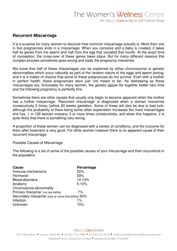
Incidence • 15-20% of all pregnancies • 11-13 % in first pregnancy • 13-17 % after first abortion • 38 % after two abortions • 55% after three abortions
Recurent Miscarriage Etiology Explained • Anatomic (Sporadic) 12%-16% • Endocrine 17%-20% • Luteal phase deficiency • Uncontrolled DM • PCOS • Immunological 10%-16% • Anti phospholipid syndrome • Environmental • Alcohol, Smoking • Genetic factors 3.5-5% Un-explained 50%
Anatomical Factors • What are the congenital & acquired • uterine anomalies leading to RSA? • How will you manage?
Uterine Abnormalities • CONGENITAL (Mullerian Duct abnormalities) • UTERINE NEOPLASMS (Growth) • IATROGENIC (Acquired)
ANATOMICAL CAUSES • Septate uterus • Intrauterine adhesions • Bicornuateut (unequal horns) • Unicornuate uterus • T shaped uterus • Submucous fibroids • Large endometrial polyps
How they affect……. • Smaller Uterine Cavities • Fewer suitable implantation sites • Aberrations of vascularisation • May be accompanied by cervical incompetence Lead to both early & later pregnancy losses
Septate Uterus • Most COMMON anomaly 55% • May be complete/ incomplete/segmental 25% early abortions 6.2% late abortions & Premature labors
Unicornuate Uterus • 20% of anomalies • Agenesis or hypoplasia of one Mullerian duct • May be alone or accompanied by Rudimentary horn With presence / absence of cavity Communicating / Non communicating • Associated Renal anomalies occur in 40% patients Ipsilateral to hypoplastic horn
Unicornuate Uterus • Abortion Rate 51%, Premature labours, malpresentations, IUGR, Uterine rupture & ectopic pregnancies common • Cervical encerclage to improve pregnancy outcome • Rudimentary Horn resected to prevent dysmenorrhoea, haematometra,ectopic pregnancy
Uterus Didelphys • Least common anomaly -5-7% • Failure of lateral fusion of uterus &vagina • Abortion rate 43%,Premature birth rate 38% • Resection of Vaginal septum if there is difficulty in intercourse / vaginal delivery • Strassmann Operation not indicated
Bicornuate Uterus • 10% of anomalies • Incomplete fusion of Uterine horns at level of fundus • Two separate but communicating endometrial cavities • Abortion rate 32% Preterm labour 21% • Strassman Metroplasty / Place IUCD in one horn
Arcuate Uterus • Near complete resorption of u-v septum • Mild concave indentation at fundus • ? Anomaly / ? Anatomic variant • Data conflicting Abortion rates ?45% ?13% • Treatment expectant
T shaped Uterus • Diethylstilbestrol treatment for Premature labour started 1940 Banned 1970 • 69% female foetuses suffered Uterine anomaly • T-Shaped uterus, small uterus, constriction rings, • Cervical hypoplasia, cervical incompetence, Anterior Cervical collar, pseudopolyps • 2 fold increase in abortion rates & 9 fold increase in Ectopic pregnancy rates
T SHAPED UTERUS- INFECTION MALA ARORA
Uterine Neoplasms • Endometrial Polyps
PERIOSTEAL ENDOMETRIAL POLYP MALA ARORA
Leiomyomas (Fibroids) most common…. 20-50% of reproductive women When will you considerfibroids responsible ?
Preconception myomectomy to improve reproductive outcome can be considered on an individual basis • It is likely to have a place only in women who have recurrent pregnancy loss, • large submucosal fibroids, and no other identifiable cause for recurrent miscarriage Ouyang DW, Obstet Gynecol Clin North Am. 2006
Iatrogenic… Intrauterine adhesions ,“Asherman’s Syndrome” • Lead to Poor implantation, • Decreased blood supply , • infection Abortion rates 40% Preterm labour 23% Management :-Hysteroscopic excision of adhesions
HYSTEROSCOPIC CORRECTION • All of the above have a good pregnancy rate post hysteroscopic correction • Except ashermans syndrome
Anatomical Factors • When will you label a patient as a case of incompetent Cervix? • What are the different surgical procedures? • Role of prophylactic surgery?
USG follow up weekly in cases of prior 2nd trimester loss • Funneling of >25% cervical length and/or <2.5 cms cervical length before 24 weeks of pregnancy • Cervical cerclage reduces the rate of preterm birth Carp et al, 2007 • Emergency cerclage: beneficial if no infection or uterine contractions
Genetic Etiology • Chromosomal 3.5%-5% • Fetal chromosomal abnormalities • Parental balanced chromosomal rearrangement • Single gene disorders • Alpha thalassemia major • Thrombophilia • X linked dominant disorders
Risk Factors for Karyotypic abnormalities Gestational age Higher in early gestation 90% in anembryonic preg/Blighted ova 50% at 8-11wk 30% at 16-19 wk 6-12% >20wk
Risk Factors for RM • Advanced maternal age • Affects ovarian function, giving rise to a decline in the number of good quality oocytes, resulting in chromosomally abnormal conceptions that rarely develop further. • RM risk -75% in women >45years • Previous number of miscarriages
Spontaneous Miscarriage • 10-15% of recognized pregnancies • Mostly sporadic ; 80% losses in 1st 12 wks • 50-70% due to chromosomal anomalies • Autosomal trisomy 50-60% • 13,16,18,21,others • Monosomy X-20% • Triploidy –15% • Tetraploidy-5% • Unbalanced translocation-3-5% Parental Karyotypes normal Minimal recurrence risk
In Recurrent Miscarriage (RM) • Fetal chromosomal abnormality in only 25-32% of product of conception (POC) • This may be due to abnormalities in the egg, sperm or both. • The most common chromosomal defects are Trisomy, Monosomy, Polyploidy • Sperm aneuploidy (13,18,21,X,Y ) directly influences the rate of aneuploidy in the conceptus (Carrell et al 2003)
In Recurrent Miscarriage • Parental chromosomal abnormality (Balanced chromosomal rearrangements) • General population 6 in 1000(0.6%) • RM 4.1-11% *3-5% of couples with RSA are carriers of balanced chromosomal rearrangements
Parental Chromosomal Abnormalities • Translocation (commonest) (1in 500) • Reciprocal [50%] • Robertsonian [24%] • Mosaicism for a numeric aberration[12%] • Inversion
Translocation Translocation is exchange of chromosomal segments between two, non-homologous chromosomes. Source-Internet
Translocations Two major types Reciprocal translocation-two non-homologous chromosomes exchange information Robertsonian translocation -two non-homologous acrocentric chromosomes break at the centromere and the long arms fuse. The short arms are often lost. Source- Emery’s book of principles of Medical Genetics
Diagnosis • Karyotype of the abortus ( fetal/placental tissue) • Peripheral blood Karyotyping of the parents in all couples with RM
Karyotype of Products of Conception • Successful culture requires healthy cells derived from the fetus • Unsuccessful in upto 50% of cases • Maternal overgrowth of fetal cells • Poor growth of abortus tissue esp. if there is a long time interval from the demise until the culture is performed • Poor chromosome morphology
Karyotype of Products of Conception • No definite recommendations for routinely obtaining abortus karyotype (ACOG 2001) • Karyotype analysis of abortus tissue for couples with a subsequent second or third pregnancy loss (Hogge, et al 2003) • If abortus is aneuploid, maternal cause is excluded (ACOG, 2001) • If POC karyotype not possible, do parental karyotype
Karyotype of Products of Conception • Normal • Abnormal (trisomy or chromosomal rearrangement) Both requires parental karyotype Direct parental karyotype is more cost effective No need for first abortion
Why Karyotype of the Parents ?? • Individuals with Balanced Chromosomal Rearrangement usually phenotypically normal • Are at risk of having conceptus with • normal • balanced phenotypically normal • unbalanced • spontaneously aborted • phenotypically malformed
Single Gene Disorders in RM • Second and 3rd trimester losses • Alpha Thalassemia • Myotonic dystrophy • X linked Dominant disorder • IncontinentiaPigmenti • Chondrodysplasiapunctata • Focal dermal hypoplasia of Goltz • Rett Syndrome • Aicardi Syndrome
Single Gene Disorders in RM • Hereditary thrombophilia • First and later trimester losses • Microthrombosis in placenta ;Impaired uteroplacental circulation • Factor V Leiden gene mutation Evidence based Prothrombin G 20210A mutation inc. risk • Protein C,S deficiency • Antithrombin III No significant association • MTHFR C677T mutation • Combination of any of above-Increased risk
Genetic Evaluation and Testing Recommendation • History of • Recurrent miscarriage • Clotting disorder • Still birth/neonatal death • Babies with dysmorphic features • Infertility • Mental retardation /developmental delay • Inherited disorder (J Gen Counsel ;14(3)2005)
Karyotypic abnormalities in couples with Recurrent abortions Dubey et al. Ind J Hum Genet 2005 • Total Couples n=742(1484 cases) • Duration -12 years • Chromosomal rearrangements = 52 (7% ) • Structural aberrations 22 (2.9%) • Reciprocal (6,8,11,18)=15 (68.2%) • Robertsonian (21,22,13,14)=4 (18.1%) • Inversion(4)=1 (9%) • Deletion=2 • Numerical anomalies (mosaics with XO,XXX, XXY)= 9 (1.2%) • Chromosomal variants (para centromeric heterochromatin/fragile sites) = 21 (3.2%)
EGG 80% Venn diagram of the responsibilities of Reproductive Failure SPERM 10% UTERUS 10%
Doubtful causes of RSA • TORCH infections • Endocrine and metabolic disease • Untreated adrenal hyperplasia, hypothyroidism & diabetes mellitus. • Exogenous causes • Environmental factors, alcohol, street drugs, anesthesia gases etc
Its time to say goodbye to TORCH tests……. Cochrane Review has categorically proven in multiple meta-analysis that none of the “TORCH” group of infections are responsible for RECURRENT SPONTANEOUS ABORTIONS TORCHTESTS
So which infections, if any are responsible for RSA? Female • Viral infections ? ? • Coxasackie B • Parovo-virus B • Bacterial infections • Bacterial Vaginosis • Tuberculosis • Chlamydia trachomatis Male factors: • Semen infections can cause anueploidy and be the reason of RSA