Download
1 / 22
240 likes | 456 Views
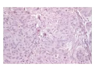
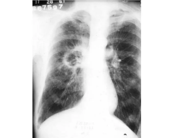
Large cell carcinoma. Accounts for 5-10% of all lung cancers. Strongly associated with cigarette smoking. The lesion occurs peripherally and grows rapidly, with early metastases and a poor outcome They lack any diagnosic features to suggest their diagnosis prior to biopsy. .

E N D
Large cell carcinoma • Accounts for 5-10% of all lung cancers. • Strongly associated with cigarette smoking. • The lesion occurs peripherally and grows rapidly, with early metastases and a poor outcome • They lack any diagnosic features to suggest their diagnosis prior to biopsy.
Pancoast tumors • Represent 1-3% of all lung cancers . • Typically involve the lower trunks of the brachial plexus, intercostal nerves, stellateganglion, adjacent ribs, and vertebrae. • More than 95% are NSCC . • Horner's syndrome, mediastinal and supraclavicular adenopathy and vertebral body invasion portends a poorer prognosis
Pancoast Tumours Imaging • MRI is more accurate in identification of the extent of tumor involvement; it is superior to CT scanning in the detection of invasion of adjacent organs (eg, vertebral bodies, brachial plexus, subclavian vessels). • CT or MRI of the brain is recommended in the initial evaluation, because distant metastases to the brain are not infrequent
Differential Diagnosis of an opacity at the Superior Sulcus • Mesothelioma. • Lymphoma. • Plasmacytoma. • Metastatic malignancies (thyroid, larynx). • Lymphomatoid granulomatosis. • Cervical rib syndrome. • Tuberculosis. • Fungal infections.
Small Cell Lung Cancer • strong association with smoking. • Rapid growth. • Early spread to distant sites. • Exquisite sensitivity to chemo and radiotherapy. • Frequent association with distinct paraneoplastic syndromes. • Surgery usually plays no role in its management, except in rare situations (<5% of patients) in which it presents at a very early stage as a solitary pulmonary nodule
Small cell lung cancer • 18% of all lung cancers. • Often present with bulky hila and mediastinal lymph node masses. • TNM system does not provide important prognostic information; only useful in <5%.
With central tumors, distinguishing primary tumor from lymph node metastasis may be impossible
International Staging System for Lung Cancer • This is the common evaluationframework,because, patient treatmentoptions and prognosis are directly related to their tumor stage at presentation . • Derived from a TNM classification scheme with four separate stage groups from I to IV. Stage I reflects the best prognosis, stage IV the worst.
Tumor (T) • TX - Positive malignant cytology, no lesion seen • T1 - Diameter smaller than or equal to 3 cm • T2 - Diameter larger than 3 cm • T3 - Extension to pleura, chest wall, diaphragm, pericardium, within 2 cm of carina, or totalatelectasis • T4 - Invasion of mediastinal organs (eg, esophagus, trachea, great vessels, heart), malignant pleural effusion, or satellite nodules within the primary lobe
T1 Tumor • Diameter of 3 cm or smaller , surrounded by lung or visceral pleura.
T2: A tumor with any of the following features: • Larger than 3 cm. • Associated with atelectasis or post-obstructive pneumonitis that does not involve the entire lung . • Invades the visceral pleura.
T3:A tumor of any size that directly invades any of the following: • The chest wall (including superior sulcus tumors), diaphragm, mediastinal pleura, parietal pericardium.
T3:A tumor of any size that directly invades any of the following • Tumor in the main bronchus less than 2 cm distal to the carina (but without involvement of the carina). • Tumor associated with atelectasis or obstructive pneumonitis of the entire lung.