Download
1 / 73
810 likes | 1.37k Views
Diagnosis, Evaluation, and Treatment of Stroke. Fariborz Khorvash, MD Assistant Professor of Neurology. Topics. Definitions Evaluation of Suspected Stroke Evaluation of TIAs Stroke Prevention Evaluation & Treatment of Ischemic Stroke Evaluation & Treatment of Hemorrhagic Stroke. Case #1.

E N D
Diagnosis, Evaluation, and Treatment of Stroke Fariborz Khorvash, MD Assistant Professor of Neurology
Topics • Definitions • Evaluation of Suspected Stroke • Evaluation of TIAs • Stroke Prevention • Evaluation & Treatment of Ischemic Stroke • Evaluation & Treatment of Hemorrhagic Stroke
Case #1 • 62 year old woman presents after a abrupt onset of blindness in her left eye while shopping today. Sx resolved en route to ER via EMS about 20 minutes after they started. • VS Afeb; BP 148/78; P 68 • Exam unremarkable in ER What’s the diagnosis?
Case #2 • 76 yo male, rehabbing at local NH after recent hip fx, has abrupt onset of slurred speech and left arm/leg weakness. • Sx persistent in ER. Pt has no complaints. • VS Afeb; BP 188/96; P 72 • Head CT is “negative” What’s the diagnosis?
Case #3 • 33 yo woman presents with “worst headache of her life”, abruptly starting 1 hour ago. • On exam, she is mildly confused, has mild nuchal rigidity, but no other focal findings • VS Afeb; BP 155/82; HR 58 What’s the diagnosis?
Classification of Stroke • 2 broad categories of stroke: • Ischemia • Inadequate blood supply (oxygen & nutrients) to an area of the brain • Hemorrhage- • Leakage of blood into the closed cranial cavity • Direct damage to tissue by compression/edema
Epidemiology of Stroke • Incidence in US • ~700K per year (~200K are recurrent) • 80-90% are ischemic • Male:Female ratio 1.25:1 • Ratio reverses after age 80 • Higher rates in Blacks, Hispanics, & Native Americans
Risk Factors • Heart disease • AFib, Valvular Dz, MI, endocarditis • Hypertension • Smoking • Diabetes/Metabolic Syndrome • Dyslipidemia • Pregnancy • Drug Abuse/Meds • Bleeding Disorders/Anticoagulant Use
Ischemic Stroke • Thrombosis • In situ arterial obstruction • Arteriosclerosis, dissection, FMD • Superimposed thrombosis • Embolism • Arterial obstruction from debris from another source • Systemic Hypoperfusion • Circulatory collapse • Multiorgan involvement
Thrombosis • Large Vessel Disease • Common & Internal Carotids • Circle of Willis & proximal branches • Small Vessel Disease • Penetrating arteries • “Lacunar Stroke” • “Stuttering” course
Embolism • Cardiac • Atrial fibrillation • Heart valves, atrial thrombus, recent MI, dilated CM, endocarditis, recent CABG • Aortic • Arterial (e.g. carotids) • Other/Unknown • DVT- “Paradoxical embolus” • Abrupt onset, rapid improvement
Hypoperfusion • Shock • Cardiogenic, septic, hypovolemic • Sx are more diffuse/nonfocal • “Border-zone regions” • Cortical blindness • Stupor • Proximal Weakness
Hemorrhagic Stroke • Intracerebral Hemorrhage (ICH) • Bleeding within the brain tissue • Forms a hematoma • Growth stopped by tamponade or leaking into the ventricles or CSF • Headache, vomiting, delirium • Progressive sx
HTN Trauma Bleeding Disorder Inherited Acquired, i.e. meds Amyloid Drug use Cocaine Amphetamines AVMs Bleeding into tumor Vasculitis Causes of ICH
Hemorrhagic Stroke • Subarachnoid Hemorrhage (SAH) • Bleeding into CSF on outer aspect of brain • Quick rise in ICP • Sudden onset headache in 97% • Aneurysm & AVMs are most common cause
Seizure with Todd’s Paralysis Syncope Migraine Head Trauma Brain tumor Metabolic Causes Hypoglycemia Hyponatremia Intoxication Uremia/ARF Hepatic Encephalopathy Conversion Disorder Differential Diagnosis
Initial Evaluation:Physical Exam • Vital signs • Temperature, Pulse, Blood Pressure • Pulses • Carotid Bruit • Cardiac Exam • Funduscopic exam • Skin exam • Signs of trauma
Initial Evaluation:Physical Exam • Neurologic Exam • Level of consciousness/GCS • Language/Speech • Cranial nerves • Vertigo, diplopia, ataxia • Visual deficits • Weakness/Paralysis • Reflexes/ Babinski
CBC with platelets Electrolytes, Bun, Cr Glucose LFTs PT/PTT O2 Sat ECG Chest XRay ESR Blood Cultures ANA Tox screen Alcohol level Blood type & cross Urine/Serum HCG Hypercoaguability Profile Initial Evaluation: Studies
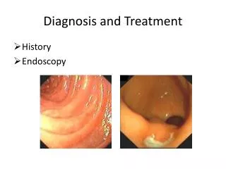
Initial Evaluation: Imaging • CT Scan • “R/O Bleed” • Sensitivity much better after 24 hrs for ischemic stroke • Early signs (<6 hrs) • May indicate worse prognosis
Initial Evaluation: Imaging • MRI • T1/T2 images, DWI • Provides immediate evaluation of ischemia • Not available for emergency use in many settings
Further Evaluation: Carotids • Carotid U/S for stenosis • If ASVD, but no stenosis… • Risk Factor Modification • If stenosis, consider… • Carotid Endarterectomy • ?Carotid Stenting • Vertigo & Syncope are not considered symptomatic
Treatment of Carotid Stenosis with Symptoms • 100% occlusion • No treatment • 70-99% occlusion • If good 5-yr survival & risks <6%, early CEA (within 2 weeks) • 50-69% • If above criteria & male, early CEA • If female, medical mgt • <50% • Medical management
Further Evaluation: Echo • Echocardiography indicated for • Patients who may need anticoagulation • Atrial fibrillation • Risk of atrial thrombus • Recent MI • Risk for Endocarditis • TEE is more sensitive than TTE, but will it change management?
Further Evaluation: Intracranial • Not necessary for all patients • Consider… • Pts <50 without a clear source • Pts with recurrent stereotyped TIAs • Posterior circulation event without cardiac source • Prior to CEA • CTA vs. MRA vs. TCD
Transient Ischemic Attack • Sudden onset of neurologic dysfunction that lasts less than 24 hrs, brought on by presumed transient ischemia to a portion of the brain • May be better to describe as sx <1 hr with no evidence of infarction • May have infarct even with sx lasting a few hours (~50% of TIA patients have MRI evidence of ischemia)
TSI? • Transient Sx Associated with Infarction • No established diagnostic criteria • In one case series, 15% of TSI pts had a recurrent stroke in-hospital vs. 0% in TIA group.
Hospitalize for TIAs? • Could consider home if able to expedite urgent outpatient work-up • AHA does not make a recommendation re: hospitalization • One study suggested cost-effective if 24-hr stroke risk is >5%
Risk of Stroke post-TIA • NASCET trial suggested 90-day stroke risk of 20% with non-retinal TIAs (higher than for true stroke) • 2000 JAMA study • 5% risk w/in 2 days • 11% risk w/in 90 days • Higher risk with age >60, DM, sx >10 min, weakness, speech impairment • 2004 Neurology study: 21% risk of stroke/MI/death within 1 year of TIA
ABCDs of TIAs • Age >60 = 1 pt • Blood Pressure >140/90 = 1 • Clinical Features • Unilateral weakness = 2 • Isolated speech deficit = 1 • Other = 0 • Duration • >60 minutes = 2 • 10-59 minutes = 1 • <10 minutes = 0 Risk of “early stroke” Score ≤ 3: 0% 4: 1-9% 5: 12% 6: 24-31%
Secondary Prevention of Stroke • Risk factor modification • Antithrombotic therapy • Anticoagulant therapy
Stroke Prevention: Risk Factors • Hypertension • Goal <130/80 • SHEP Study, ISH in pts >60 • Dropped SBP from 155 to 143 • 36% reduction in stroke over 4 years • Pts >80 may not benefit as much & aggressive BP lowering may increase mortality • Diuretic +/- ACEI as 1st line
Stroke Prevention: Risk Factors • Smoking • Stop it • Diabetes • Goal A1c <7, i.e. normoglycemic • Metabolic syndrome
Stroke Prevention: Risk Factors • Dyslipidemia • Evidence not as strong as may think, but still a good idea, especially given other vascular disease • SPARCL Study • Atorvastatin 80 mg/day in pts 1-6 months from CVA/TIA • Mean LDL reduction 56 • Endpoint was stroke: 16% RRR, but only 2.2% ARR (NNT ~50)
Stroke Prevention: Risk Factors • Dyslipidemia, continued • For average-risk patient, goal LDL <100 • For high-risk, goal <70 • Diabetes • Prior CAD • Multiple RFs with continued smoking
Stroke Prevention: Risk Factors • Lifestyle Modification • Weight loss • Exercise • Dietary changes • Reduce alcohol intake, especially heavy drinkers • ?Homocysteine • Consider B12, B6, Folate (MVI doses OK)
Antiplatelet Therapy • Aspirin • 20-25% reduction in stroke (& MI or other vascular death) • Standard doses of 81-325 mg as good as higher doses • 81 mg dose just as good and less risk of bleeding • ASA-non-responders?
Antiplatelet Therapy • Clopidogrel (Plavix) • 8% RRR vs. ASA for stroke/MI/Vasc death • 5.3% vs. 5.8%: NNT ~200 • All for only $100+/month • ?2nd-line therapy or ASA-allergic patients • No increased bleeding vs. ASA, but combo should be avoided • No neutropenia (like ticlopidine)
Antiplatelet Therapy • Dipyridamole • Alone 50-100 mg TID • Aggrenox (200mg ER-DP & 25mg ASA) BID • 2 studies have shown ~3% ARR (NNT 33) over ASA alone for stroke prevention • Some guidelines are suggesting this a 1st line therapy over ASA alone for stroke prevention • Cost >$100/month
Anticoagulant Therapy • Warfarin has only been proven effective in primary prevention of stroke in the setting of atrial fibrillation • AF is responsible for 1/6th of all strokes in patients older than 60 • Risk reduction • Warfarin about 3 times as effective as ASA • Absolute annual risk reduction of ~3% • “Low Risk” patients may consider ASA rx
Risk Stratification for Stroke • Highest Risk: Prior Stroke or TIA • High Risk: Any of the following • Prior thromboembolism • Female >75 yo • SBP >160 • Heart failure/LV dysfunction • Moderate Risk: None of above, but HTN • Low Risk: None of the above, no HTN
Choice of Medication Based on SPAF-III Trial, Lancet 1996
Treatment of Ischemic Stroke • Thrombolysis • Blood Pressure Management • Antithrombotic Therapy • Management of Medical Complications