Download
1 / 29
300 likes | 348 Views
Encephalitis is brain inflammation usually caused by viral infections like Herpes simplex. Dr. Alka Stoelinga explains its etiology, clinical features, examinations, investigations, management, prognosis, and complications. Additionally, learn about cerebral malaria, its pathogenesis, clinical features, examination, and investigation.

E N D
ENCEPHALITIS,CEREBRAL EDEMA Dr. Alka Stoelinga
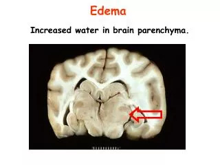
Encephalitis means inflammation of brain parenchyma • Cause: usually viral • Brain inflammation also develops in bacterial and fungal meningitis • Not an isolated entity • Usually associated with meningeal inflammation together • Also term as “Meningoencephalitis” • Commonest virus is Herpes simplex which reaches brain via the Olfactory nerve • Meningism is the triad of • Nuchal rigidity (neck stiffness) • Photophobia (intolerance of bright light) • Headache. Dr. Alka Stoelinga
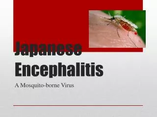
Etiology: • Arbo virus • Japanese B encephalitis • Herpes simplex • Coxsackie • Epstein-Barr viruses • Adenovirus • Varicella zoster • Influenza • Measles • Entero virus • Mumps (Aseptic meningitis) • HIV • Rabies Dr. Alka Stoelinga
Clinical features: • Acute onset of Fever, Headache, Meningism and Drowsiness in mild cases. • In severe illness: focal signs(e.g Aphasia, hemiplegia or cranial nerve palsies), siezures and coma may develop • Coma: Rapid deterioration in sensorium • Altered consciousness, focal signs and seizure are more common in encephalitis as compared to meningitis • Patient may agitate(while in meningitis patients is usually calm, conscious, drowsy and non-agitated) Dr. Alka Stoelinga
Examination: • Coma/Altered level of consciousness • Meningeal signs may be present • Focal neurological deficits • Hypertonia/Brisk reflexes/Extensor plantar • Opthalmoscopic examination: Look for papilloedema Dr. Alka Stoelinga
Investigation: • CT scan head :Shows area of edema • In Herpes simplex encephalitis CT scan show low density lesions in the temporal lobes • MRI head • EEG : Shows slow wave changes • CSF analysis • Cell: 10-2000 • Mainly Lymphocytes • Glucose usually normal • Protein normal or Slightly raised 5. Viral serology of blood and CSF 6. PCR: Herpes simplex virus DNA can be detected by PCR Dr. Alka Stoelinga
Management: • Supportive • Fluid balance • Control of seizure • Treatment of raised ICP:Dexamethasone 4mg QID for raised intracranial pressure • Herpes simplex encephalitis: Acyclovir • Dose: 10mg/kg/dose IV 10 hourly • Acyclovir should be given to all suspected cases of viral encephalitis • Anticonvulsants if presence of Epilepsy Dr. Alka Stoelinga
Prognosis: • Mortality is 10-30% when antiviral drugs are used, without antiviral mortality is 70% • Survivors may have cognitive impairment or developed epilepsy Complications: • Focal Neurological deficit • Loss of cognitive function • Seizure Dr. Alka Stoelinga
CEREBRAL MALARIA • Severe form of falciparum malaria • Common in tropical area • Treatable condition Pathogenesis : • Sequestration of parasitized RBC into blood vessels of internal organs: Brain • Rouleax formation • Cerebral anoxia • Hemolysis Dr. Alka Stoelinga
Clinical features: • Chills, Persistent high fever, headache, orthostatic hypotension, myalgia • Red blood cell (RBC) sludging that leads to capillary blockage at several sites • The three initial stages of Cerebral Malaria are: • Cold Stage: It ranges from chills to extreme shaking for 1-2 hours • Hot Stage: It is characterized by a high fever up to 107°F (41.7°C) for 3-4 hours • Wet Stage: It is characterized by profuse sweating for 2-4 hours • There are three primary symptoms of cerebral malaria which are common in both adults and children: • Impaired consciousness with non-specific fever, • Generalized convulsions and neurological abnormalities, and • Coma that lasts for 24-72 hours, initially rousable and then unrousable Dr. Alka Stoelinga
It also manifests with • signs of increased intracranial pressure • Hemiplegia • Encephalopathy • Delirium • seizures and • Coma • If not treated on time, it can lead to complications like • Jaundice • Hemoglobinuria • a tender and enlarged spleen • acute renal failure • Uremia Dr. Alka Stoelinga
Examination: • Altered consciousness • Pallor • Icterus • Bleeding manifestation • No focal neurological deficit • Splenomegaly Dr. Alka Stoelinga
Investigation: • Peripheral blood smear: • Thick and Thin smear • CSF analysis • Blood Sugar, electrolytes, ABG, urea, creatinine Dr. Alka Stoelinga
INTRACRANIAL SPACE OCCUPYING LESION(ICSOL) Dr. Alka Stoelinga
TUBERCULOMA (TBM) • Non-neoplastic mass • Tumor like growth of Tuberculous tissue in the central nervous system, characterized by symptoms of an expanding cerebral, Cerebellar, or spinal mass • Presents as ring enhancing lesion • Seizure: Partial seizure • Focal neurological deficit • Headache Dr. Alka Stoelinga
Investigation: • CT scan head: Ring enhancing lesion • AFB smear • Mantoux • CXR Treatment: • Similar to that of tubercular meningitis Dr. Alka Stoelinga
NEUROCYSTICERCOSIS (NCC) • Cysticercosis, is an infection which results from the ingestion of the eggs of the pork tapeworm (uncooked/undercooked pork meat), Taenia solium • Neurocysticercosis, is when the brain or spinal cord (CNS) is affected by the larval stage of T. solium • Neurocysticercosis is the most common helminthic (tapeworm) infestation to affect the CNS worldwide • Is the prime cause of acquired epilepsy. Dr. Alka Stoelinga
Clinical features: • Seizures: Partial/ Generalised • Hydrocephalus • Features of raised ICP • Stroke, focal neurological deficits • Meningitis Dr. Alka Stoelinga
Investigations: • Neuroimaging: • MRI(early stage)/CT head(late phase) • Ring enhancing lesion • Scolex • Calcification • Perilesional edema 2. Fundoscopy 3. Soft tissue X-ray 4. Serologic test: ELISA Dr. Alka Stoelinga
Treatment: • Treatment of seizure Antiepileptics • Treatment of Hydrocephalus if present • Cysticidal therapy • Indicated for active lesions • Albendazole 15mg/kg/d for 8-28 days • Praziquantel • In immature cyst stage High dosage of corticosteroids • In the colloid cyst stage Surgical removal of the cyst, along with albendazole is indicated • In calcified dead cysts No treatment Dr. Alka Stoelinga
TBM NCC Due to ingestion of T.solium egg Small size <10mm and are multiple in number Usually situated in the periphery with well defined margins Less perilesional edema Calcification Hydrocephalus is uncommon • Secondary to primary or post primary TB • Bigger size than NCC • Situated in the midline and margins are thick and irregular • More perilesional edema seen • Calcification • Midline shift and hydrocephalus Dr. Alka Stoelinga
BRAIN ABSCESS Predisposing factors: • Hematogenous spread: Can occur from any site of primary infection but is commonly associated with: • Infection of chest, including lung abscess, bronchiectasis and empyema • Infection of heart such as infective endocarditis • Infection of bone or dental abscess can be primary site of infection • Direct/Local spread: Penetrating injury of skull, PNS, Middle ear • Mostly involve Frontal and temporal lobes Dr. Alka Stoelinga
Causative organisms: • Mixed aerobic and anaerobic organisms are involved • Staph aureus • Streptococcus milleri • Enterobacteriae Dr. Alka Stoelinga
Clinical feature: • Illness develops more gradually than acute bacterial meningitis. • Fever, headache, drowsiness, altered sensorium • Partial seizure • Focal neurological deficit • Features of meningitis • Septicemia Dr. Alka Stoelinga
Investigations: • Neuroimaging: CT scan head • Single or multiple ring enhancing lesion with perilesional edema(central low density and surrounding edema) • CSF analysis usually not helpful and C/I if there is evidence of raised ICP • Blood : TC,DC,ESR,C/S Dr. Alka Stoelinga
Treatment: • Emperic antibiotic therapy must be started • Inj Benzyl penicillin 2 million units IV hourly +Inj Chloramphenicol 1-2 gm IV 6 hourly + Inj Metronidazole 500mg IV 6 hourly • Steroid: Inj Dexamethasone 4-25 mg 6 hourly followed by tappering of dose to reduce cerebral edema • Along with drainage of Pus • 3rd generation Cephalosporin+Metronidazole+Cloxacillin/Vancomycin • Antibiotics should be continued for 6-8 weeks. Dr. Alka Stoelinga