Download
1 / 14
160 likes | 498 Views
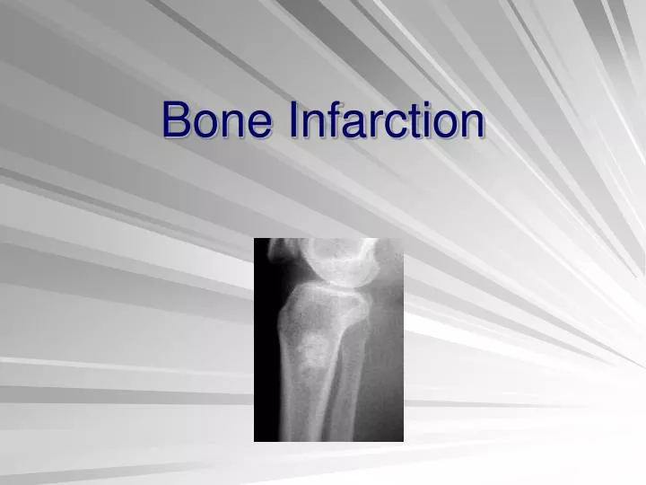
Bone Infarction. Case “Inspiration”. Sambeaux Brubaker 5.5 year old MC Rottweiler Left TTO surgery 7/10/08 Post-op recheck 8/5/08—not progressing as well as hoped. Left stifle/tibia radiographs made Increased mineral opacity in tibial medullary cavity Differential diagnoses: Bone infarct

E N D
Case “Inspiration” • Sambeaux Brubaker • 5.5 year old MC Rottweiler • Left TTO surgery 7/10/08 • Post-op recheck 8/5/08—not progressing as well as hoped
Left stifle/tibia radiographs made • Increased mineral opacity in tibial medullary cavity • Differential diagnoses: • Bone infarct • Indolent osteomyelitis • Panosteitis
Causes of Bone Infarction • Trauma (surgery e.g.-THR) • Idiopathic • Small breed terrier/Sheltie dogs • Humans—Hepatic lipidosis, Sickle cell disease, chronic steroid administration, pancreatitis Thrall, 2007, Sebestyen, et al, 2000, and emedicine.com
Other Differential Dx • Metastatic neoplasia • Lymphoma • Panosteitis • Infection • Osteopetrosis/osteosclerosis Thrall, 2007 and Marcellin-Little et al, 1999
Bone Infarction • Time course • Initial signs non-specific, visible within a few months (3-6 mos)—loss of trabeculae, smooth periosteal rxn, irregular increase in opacity of medulla • More chronic—pathognomonic changes recognizable at 1 year—irregular, serpiginous, radiopaque lines in medullary cavity • “Smoke in the chimney” Sebestyen, et al, 2000
Bone Infarction • Prevalence • Uncommon, assc with THR • 14% of femurs with femoral prostheses (15/110) had infarcts in 2000 study • 8/53 (15%) had uncemented THR • 7/57 (12%) had cemented THR Sebesten, et al, 2000
Histopathology—what’s really going on in there? • Ischemic osteonecrosis • Center (radiolucent)—dead marrow and dead osteocytes, empty lacunae • Periphery (radiopaque)—dystrophic mineralization • Periosteal reaction—woven bone • Different from endosteal reaction, which is live bone proliferation! Sebestyen, et al, 2000
Pathophysiology • Trauma to nutrient artery • Nutrient artery relatively more proximal in dogs than humanstrauma during THR • Younger dogs may be more affected because nutrient artery provides most of blood flow (older dogs use more periosteal vessels) • No clinical signs, but possibility of malignant transformation Sebestyen, et al, 2000
Malignant Transformation • Humans—osteosarcoma, fibrosarcoma associated with infarcted sites • 1999 report of dog with bilateral femoral infarcts—OSA developed unilaterally 5 yrs post-op • Track patients especially well if they develop infarcts! Marcellin-Little, et al, 1999
References • Marcellin-Little DJ, DeYoung DJ, Thrall ED, and Merrill CL. Osteosarcoma at the site of bone infarction associated with total hip arthroplasty in a dog. Veterinary Surgery 28: 54-60. 1999. • Sebestyen P, Marcellin-Little DJ, and DeYoung BA. Femoral medullary infarction secondary to canine total hip arthroplasty. Veterinary Surgery 29: 227-236. 2000. • Textbook of Veterinary Radiology, 5th ed. Thrall DE, ed. 2007. pp 306, 309. • www.emedicine.com “Bone infarction”