Download
1 / 31
310 likes | 355 Views
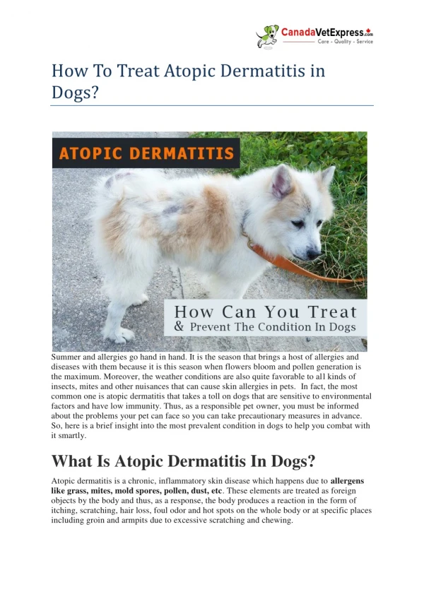
Explore a case study of a 64-year-old male with a rapidly growing conjunctival mass in the context of atopic dermatitis. Learn about differential diagnosis, biopsy, treatment strategies, and critical features to consider. Discover risk factors and connections between atopic dermatitis and squamous cell carcinoma.

E N D
Conjunctival mass in atopic dermatitis Nora V. Laver, MD Tufts Medical Center New England Eye Center Tufts University School of Medicine Boston University Medical Center
Clinical History NewLeftConjunctivalMass • 64 year old male presented forevaluationofamassonconjunctivaOS. • Rapidgrowthsincefirstnotedtwo months earlier. • FBS&increasedphotosensitivityOS
MedicalHistory • • SHx: MHx: • Formersmoker:1.5ppdx41years Chewedtobaccox10yearsFormerEtOH,NoDrugs Retiredmilitaryofficer,chemicalexposure • HTN • HLD • DM • Asthma • Vascularhamartoma • Rosacea • • • • Medications: • • • • • QvarVentolinHFADiovan ProtopicUngOUPFBIDOU • OcularHx: • SevereocularallergieswithatopicblepharitisOU CornealneovascularizationOU • CataractOU • Allergies: no known drug allergies,environmentalallergies •
OcularExam • • VA:CC20/70OD,OS20/100OD.(PHNIOU) Pupils:unabletoassessduetophotosensitivity EOM:fullOD,restrictionalldirectionsOS Lids:edema&erythema,thickkeratinizedlidmarginswithcrusting. Completemadarosis, cicatricialectropionOU Conj:1+diffuseinjwithforeshortenedfornixOU. • • OS: Largeelevatedsessilemasstemporallyw/extensionontocorneab/t2-5o’clock. • • Cornea:anteriorstromalhazewNV360OU AC:DeepandQuietIris:WNL Lens2+NSDFE:WNL • • • • 7
DDx:NeoplasticDisordersoftheConjunctiva • EpithelialLesions: • Vascular • SquamousCellCarcinoma • MucoepidermoidCarcinoma • SpindleCellCarcinoma • GlandularTumors: • Oncocytoma • SebaceousGlandCarcinoma • NeuroectodermalTumors • Melanoma • Leiomyosarcoma • Neurogenic: • Neuroma/neurofibroma • BenignEpithelialTumors:papilloma • • • HemangiomaPyogenicgranulomaKaposi’sSarcoma • ConjunctivalIntraepithelialNeoplasia(CIN) • Lymphatic: • • • Lymphangiectasialymphhyperplasialymphoma • MetastaticTumor: • Fromelsewhereintheeye(uvea,orbit,paranasalsinuses) • Breast,Lung,Kidney 14
Nowwhat?… • Otherexamfindingstolookfor? • Concerningfeatures: • Elevatedlesions • Extensivepigmentation • Fixedtounderlyingtissue • Feedervessel • PalpableLNs • Biopsy? • incisionalorexcisional? • Imaging? 16
ClinicalCourse IncisionalLeftConj.Biopsy 17
ClinicalCourse IncisionalLeftConj.Biopsy • Invasivesquamouscellcarcinoma,moderatelytowelldifferentiated. • Extendingto the edgesofbiopsysample • HPV- Subsequently,plannedforsurgicalexcision 17
ClinicalCourseContinued… • A month after the biopsy massnotedtohavegreatlyenlarged. • Pre-auricular lymphnodenoted • CToffaceandorbitswithcontrastordered. 2 5
CTFacialBoneswithContrast 2.3cm2.5cmleft parotidmass Axial image shows hyperdense soft tissue lateral conjunctiva mass abutiing the lacrimal gland (1 x 1.7 x 2.3 cm)
ClinicalCourseContinued… • Mass parotic underwentFNA. • SCCmetastaticfromtheeye. • PETscanobtained: 27
ClinicalCourseContinued… • Underwentpartialexcisionofconjunctivalmassw/applicationof5-FU • InvasiveSCC,extendingtoedgesofsamplewithlympho-vascularinvasion • Parotidectomywithneckdissection. • SCCwith4/10periparotidLN+&1/21cervicalLN+. 28
Clinicalcoursecontinued…. • ExenterationOS ModeratelydifferentiatedSCCextendingintoconj,cornea,scleraandorbitaltissues. UnderwentRadiationtoorbit,parotidandneck Initiallydidwell,butnoticednewneckmasses 10 months after initial presentation and 3 months after end of radiation treatment
CTHeadandNeck 4.6 x 3.7 x 4 cm enhancing soft tissue mass in the left neck in the area of the thyroid cartilage
Clinicalcoursecontinued…. • Underwentradicalneckdissection: • Path: poorly differentiated SCCA involving muscle, fibroadiposetissue and jugular vein. Extensiveperineuralinvasionwith3/19LN+ • • Initiallydidwell,butwasadmittedtotheOncologyserviceforfailuretothrive. • Imagingatthattimeshowed: • HilarLNnotedonrepeatCTandbonymetsinC4andamassencasingthecarotid. • Patientexpiredwithinafewdays. 31
HowfrequentlydoesCSCCmetastasize? Initialsiteofmetastasisincludedtheparotidgland,submandibularandsubmaxillaryglands,preauricular,cervicallymphnodes,lungs,andbone. 32
RiskFactorsforRecurrentorMetastaticCSCC • Largesize(>5-10mm) • Positivebiopsymargins • Highproliferationindexonpathology • Johnsonetal,notedaparticularlyhighrateofmet.CSCC: • Invasionintoglobe-poorprognosticfactor • Longerthanaveragedelayinpresentation(>6mo) • 10%hadasecondprimarytumor(geneticpredisposition) • Highrateofchronicocularinfection(trachoma) WhataboutAtopicDermatitis? 33
Associationb/tatopicdermatitis&SCC? • Jensenetal:incidenceratioof2.48forSCCandatopicdermatitis Cheng,etal.OR1.58ofSCCinatopicdermatitis Heinzetal,acaseseriesof6patientswithocularatopiceczemawithSCCarisingfromtheaffectedarea: • • 34
Pathophysiologyofatopyandtumorgenesis • Highratesofimmunesuppressantmedicationsinatopicpatients MayinvolveTcelldysregulation: • • AtopicEczemadisturbanceofTcellmaturation • HighincidenceofCSCCinHIV(downregulationofTcells) • IgEinterfereswith mononuclear cell tumorkillingcapacity • Inflammatorymediators(MMPs,O2free radicals)wellrecognizedasariskfactorforlungcancerinpatientswithasthma 35
Learningpoints • MetastaticCSCCisararebutdeadlydisease • Delayinpresentationisthebiggestriskfactorforvisionandlife-threateningconjunctivalmalignancy • Higherlevelofvigilancemayberequiredinpatientswithchronicinflammatoryconditions,includingatopy 36
Selected References 37 • McKelvie, Daniell M, McNab A, Loughnan M, Santamaria JD. Squamous cell carcinoma of the conjunctiva: a series of 26 cases. Br J Ophthalmol 2002; 86:168–173. • Iliff WJ, Marback R, Green WR. Invasive Squamous Cell Carcinoma of the Conjunctiva. Arch Ophthalmol 1975; 93 (2): 119-122. • Jensen AO, et al. Atopic Dermatitis and Risk of Skin Cancer A Danish Nationwide Cohort Study (1977–2006). Am J Clin Dermatol 2012; 13 (1): 29-36. • Cho JD, David DMR, Wetter DA, Bartley AC, Brewer JD. Association between atopic dermatitis and squamous cell carcinoma: a case-control study. International J Dermatol 2017; 57(3): 313-316. • Kao AA. Clinicopathologic Correlation of Ocular Surface Squamous Neoplasms at Bascom Palmer Eye Institute: 2001 to 2010. Ophthalmology 2012; 119:1773–1776. • Cheng J, et al. History of Allergy and Atopic Dermatitis in Relation to Squamous Cell and Basal Cell Carcinoma of the Skin Cancer. Epidemiol Biomarkers Prev. 2015; 24(4): 749–754. • Flynn TH, et al. Ocular surface squamous neoplasia in an immunosuppressed patient with atopic Keratoconjunctivitis. Int Ophthalmol 2012; 32:471–473. • Galor A, Karp CL, Oellers P, Kao AA, Abdelaziz A, Feuer W, Dubovy SR. Predictors of Ocular Surface Squamous Neoplasia Recurrence after Excisional Surgery, Ophthalmology 2012; 119 (10): 1974-1981. • Ramberg I, et al. Squamous cell dysplasia and carcinoma of the conjunctiva. A nationwide, retrospective, epidemiological study of Danish patients. Acta Ophthalmol. 2015; 93: 663–666 • Yousef YA, Finger PT. Squamous Carcinoma and Dysplasia of the Conjunctiva and Cornea: An Analysis of 101 Cases. Ophthalmology 2012; 119 (2):233-240. • Heinz C, et al. Squamous Cell Carcinoma of the Conjunctiva in Patients with Atopic Eczema. • Cornea 2003; 22(2): 135–137. • Shields CL, SHields JA. Tumors of the Conjunctiva and Cornea. Survey of Ophthalmology 2004; 49 (1): 3-24. • Shields JA, Shields CL, Gunduz K, et al. Intraocular invasion of conjunctival squamous cell carcinoma in five patients. The 1998 Pan American Lecture. Ophthal Plastic Reconstruct Surg 1999; 15:153–160.
The End 38 Rockport, MA