Download
1 / 15
150 likes | 300 Views
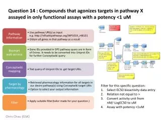
Perfusion MRI in GSK Study. Units. Perfusion is measured in mL/100g of tissue/min, or units of CBF Normal human gray matter is perfused at a rate of 50-60mL/100g/min. Measurable Parameters.

E N D
Units • Perfusion is measured in mL/100g of tissue/min, or units of CBF • Normal human gray matter is perfused at a rate of 50-60mL/100g/min
Measurable Parameters • CBF – Cerebral Blood Flow – The amount of blood moving through a given amount of tissue per unit time (can be absolute: aCBF, or relative: rCBF) • CBV – Cerebral Blood Volume – The amount of blood in a given amount of tissue at any time • MTT – Mean Transit Time – The average amount of time it takes any water molecule or particle of contrast agen to pass through the voxel vasculature
MRI Methods • Bolus Tracking - use of a contrast agent such as gadolinium as a tracer • Arterial Spin Labeling (ASL) - labels the water molecules of blood magnetically to use as a tracer
Arterial Spin Labeling Method • Tracer is a magnetic label applied to the water molecules in the blood • Magnetic label is produced by saturating or inverting the longitudinal component of the MR signal • The tagged water will pass into brain tissue and alter it’s longitudinal magnetization • A control image where no tagging is done is collected after each tagged image
Calculating CBF DM = Mcontrol – Mtag A difference image is created from the control and tag image that is proportional to CBF.
Surround Subtraction • Perfusion images are calculated from a combination of the control and tagged images • Odd indexed images are tagged • Even indexed images are controls • Perfusion weighted time series = {img[1] – (img[0]+img[2])/2}, {(img[1]+img[3])/2-img[2]}, …
Problems with Pulsed ASL • Slice profile effect alters the quality of the subtraction • Measurements are sensitive to the transit time of blood water between labeling and imaging sites
Solution • Add one or more spatial saturation pulse during the inversion time to saturate either the imaging slice or the tagging region. This is referred to as quantitative imaging of perfusion using a single subtraction (QUIPSS and QUIPSSII)
QUIPSS RF Pulse Sequence • Slice selective in-plane presaturation applied to imaging slice • Inversion tag is applied • Second saturation pulse is applied to tagging region after a delay, TI1 (done only in QUIPSSII) • After additional delay, at time TI2, an image is acquired using single shot EPI after the inversion tag
GSK Study Basic Info • 3 Study Days: A – Placebo, B – Ketamine, C – Ketamine&Lamotragine • 2 Perfusion runs ~ 5minutes long • One before the bolus and one after the bolus • One Proton Density scan acquired
GSK Study Run Breakdown • Localizer • 3D T1 • T1 flash • Fixation • VOD • VOD • VOD • Fingertap • Proton Density • Perfusion • Fixation (bolus delivered) • VOD • VOD • VOD • Perfusion
Output Images in Analysis • dM – difference image (relative CBF) • BOLD • CBF (absolute CBF is calculated if Proton Density image was specified)