Download
1 / 57
570 likes | 589 Views
Learn about the study of electromagnetic radiation interactions, electronic excitation, molecular vibrations, and nuclear spin orientations in UV, IR, NMR, and CD spectroscopy. Understand the principles, applications, and types of analysis involved.

E N D
UV, IR, NMR, CD Isariya Techatanawat, PhDDirector of Bioequivalence Study Group, Research and Development Institute, The Government Pharmaceutical Organization
Spectroscopy • Study of interaction of electromagnetic radiation. • Interaction might give rise to electronic excitations, (e.g. UV), molecular vibrations (e.g. IR) or nuclear spin orientations (e.g. NMR).
Spectroscopy • When a beam of white light strikes a triangular prism it is separated into its various components. This is known as a spectrum.
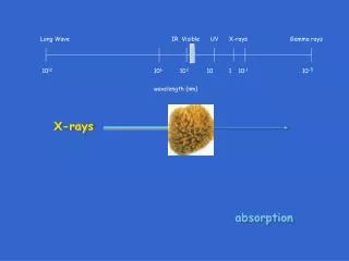
Spectroscopy • There are many other forms of light which are not visible to the human eye and spectroscopy is extended to cover all these.
Ultraviolet and Visible Spectroscopy • Ultraviolet (UV) &visible radiation comprise only a small part of electromagnetic spectrum.
Ultraviolet and Visible Spectroscopy • Wavelength: Distance between adjacent peaks (or troughs). • Frequency: Number of wave cycles that travel past a fixed point per unit of time [cycles per second, or hertz (Hz)].
Some Natural Organic Pigments • Colored compounds is a system of extensively conjugated pi-electrons.
Energy Associated with Electromagnetic Radiation • E = hν where E = energy (in joules), h = Planck’s constant (6.62×10⁻³⁴Js) ν = frequency (in seconds).
UV/Vis Absorbance • Proteins absorb at 280 nm due to presence of amino acids with aromatic rings. • Proteins absorb at 200 nm due to peptide bonds.
Infrared Spectroscopy (IR) • Absorption of infrared radiation brings about changes in molecular vibrations within molecules and 'measurements' of the ways in which bonds vibrate gives rise to infrared spectroscopy.
Infrared Spectroscopy (IR) • Atom size, bond length and bond strength vary in molecules and so the frequency at which a particular bond absorbs infrared radiation will be different over a range of bonds and modes of vibration.
The Different Types of Bonds • An organic molecule may contain quite a number of different bonds. All of these bonds will be vibrating, and clearly, different bonds will be vibrating at different frequencies. • There are two basic modes of vibration – ‘stretching’ and ‘bending’.
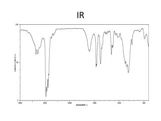
Infrared Spectrometer • Infrared spectrometer analyses compound by passing infrared radiation, over a range of different frequencies, through a sample and measuring the absorptions made by each type of bond in the compound. • This produces a spectrum, normally a ‘plot’ of % transmittance against wavenumber.
Infrared Spectrometer • Since no 2 organic compounds have the same IR spectrum, a compound can be identified with certainty by comparing its spectrum with that of a known pure compound. • If they are identical, then they are one and the same.
Nuclear Magnetic Resonance (NMR) • When some atoms are placed in a strong magnetic field, their nuclei behave like tiny bar magnets aligning themselves with the field. • Electrons behave like this too, and for this reason both electrons and nuclei are said to possess “spin”. • Any spinning electric charge has an associated magnetic field.
NMR • Just as electrons with opposite spin pair up with each other, a similar thing happens with protons and neutrons in the nucleus.
NMR • If a nucleus has an even number of protons and neutrons (e.g. 12C), their magnetic fields cancel each other out and there is no overall magnetic field. • If the number of protons and neutrons is odd (e.g.13C and 1H ), the nucleus has a magnetic field.
NMR • If the substance is placed in external magnetic field, nuclear magnet lines up with the field, in the same way as a compass needle lines up with a magnetic field.
NMR • NMR is particularly useful in the identification of the positions of hydrogen atoms (1H) in molecules.
1H NMR spectrum Ethyl benzene, C6H5CH2CH3
2D NMR • 1D protein spectra are too complex for interpretation as most of the signals overlap heavily. • By introduction of additional spectral dimensions, these spectra are simplified and some extra information is obtained.
13C NMR • 13C has only about 1.1% natural abundance • 12C does not exhibit NMR behavior. • Magnetic moment of 13C nucleus is much weaker than that of a proton. NMR signals from 13C nuclei are much weaker than proton signals. • Chemical shift range is normally 0 to 220 ppm. • Chemical shifts are measured with respect to tetramethylsilane (TMS), (CH3)4Si.
Circular Dichroism (CD) • Difference in absorption of left-handed circularly polarised light and right-handed circularly polarised light • Occurs when molecule contains one or more chiral chromophores.
Circular Dichroism (CD) • Circular dichroism = ΔA(λ) = A(λ)LCPL ‐ A(λ)RCPL • where λ is the wavelength LCPL = Left-handed circularly polarised light RCPL = Right-handed circularly polarised light
Circular Dichroism (CD) • CD of molecules is measured over a range of wavelengths. • Use to study chiral molecules. • Analyse the secondary structure or conformation of macromolecules, particularly proteins.
Circular Dichroism (CD) • Observe how secondary structure changes with environmental conditions or on interaction with other molecules. • Measurements carried out in the visible and ultra-violet region.
Circular Dichroism (CD) • Molecule contains chiral chromophores then one CPL state will be absorbed to a greater extent than the other. • CD signal over the corresponding wavelengths will be non-zero.