Download

1 / 19
190 likes | 205 Views
Explore the real-world applications of DNA fingerprinting in crime scenes, human and animal relatedness, paternity, anthropology, disease identification and more. Learn about DNA restriction enzymes and their role in cutting DNA sequences for analysis. Discover the process of DNA digestion, gel electrophoresis, and fragment analysis.

E N D
Forensic DNA Fingerprinting: Using Restriction Enzymes
DNA FingerprintingReal WorldApplications • Crime scene • Human relatedness • Paternity • Animal relatedness • Anthropology studies • Disease-causing organisms • Food identification • Human remains • Monitoring transplants
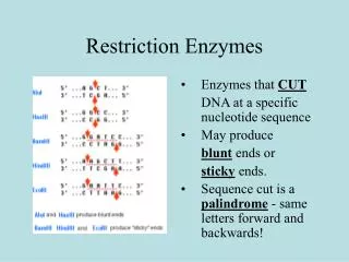
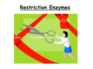
DNA Restriction Enzymes • Evolved by bacteria to protect against viral DNA infection • Endonucleases = cleave within DNA strands • Over 3,000 known enzymes
Enzyme Site Recognition Restriction site Palindrome • Each enzyme digests (cuts) DNA at a specific sequence = restriction site • Enzymes recognize 4- or 6- base pair, palindromic sequences (eg GAATTC) Fragment 2 Fragment 1
5 vs 3 Prime Overhang Enzyme cuts • Generates 5 prime overhang
Common Restriction Enzymes EcoRI – Eschericha coli – 5 prime overhang Pstl – Providencia stuartii – 3 prime overhang
The DNA DigestionReaction • Restriction Buffer provides optimal conditions • NaCI provides the correct ionic strength • Tris-HCI provides the proper pH • Mg2+ is an enzyme co-factor
DNA DigestionTemperature Why incubate at 37°C? • Body temperature is optimal for these and most other enzymes What happens if the temperature is too hot or cool? • Too hot = enzyme may be denatured (killed) • Too cool = enzyme activity lowered, requiring longer digestion time
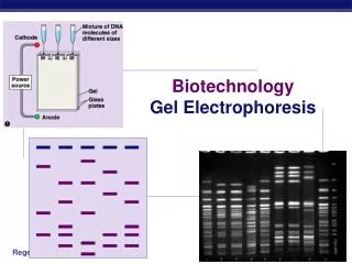
AgaroseElectrophoresisLoading • Electrical current carries negatively-charged DNA through gel towards positive (red) electrode Buffer Dyes Agarose gel Power Supply
AgaroseElectrophoresisRunning • Agarose gel sieves DNA fragments according to size – Small fragments move farther than large fragments Gel running Power Supply
Analysis of Stained Gel Determine restriction fragment sizes • Create standard curve using DNA marker • Measure distance traveled by restriction fragments • Determine size of DNA fragments Identify the related samples
PstI EcoRI Restriction Fragment Length PolymorphismRFLP GAATTC GTTAAC CTGCAG GAGCTC Allele 1 1 2 3 CGGCAG GCGCTC GAATTC GTTAAC Allele 2 3 Fragment 1+2 Different Base Pairs No restriction site M A-1 A-2 Electrophoresis of restriction fragments M: Marker A-1: Allele 1 Fragments A-2: Allele 2 Fragments +
Molecular Weight Determination Fingerprinting Standard Curve: Semi-log Size (bp) Distance (mm) 23,000 11.0 9,400 13.0 6,500 15.0 4,400 18.0 2,300 23.0 2,000 24.0