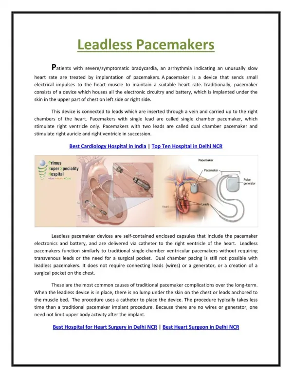
Download
1 / 15
170 likes | 429 Views
MRI in patients with pacemakers. DR PRADEEP SREEKUMAR. Types of electromagnetic fields generated by MRI. Static magnetic field-non varying ,always present around an MRI machine Gradient magnetic field-low frequency pulsed time varying magnetic fields. RF field-pulsed radio frequency field.

E N D
MRI in patients with pacemakers DR PRADEEP SREEKUMAR
Types of electromagnetic fields generated by MRI Static magnetic field-non varying ,always present around an MRI machine Gradient magnetic field-low frequency pulsed time varying magnetic fields. RF field-pulsed radio frequency field
Most MRI scanners use 1.5 to 3 tesla Equivalent to 30,000 to 60,000 times strength of earth’s magnetic field. It attracts ferromagnetic substances into the magnet “Ferromagnetic” means any substance that experiences an attractive force in presence of a magnetic field
Potential Hazards Movement and torque Electrical current induction Tissue injury by heating: increased capture thresholds Electromagnetic interference: may result in inhibition of pacing Change of reed switch state
Magnetic field interactions: vibration ,torque Induced stimulation: stimulation due to currents induced b gradient magnetic field Lead electrode heating:RF fields induce voltages in leads that may cause heating. Heating may produce tissue damage
MRI should not be done in • Newly implanted leads(<6 weeks) • Abandoned or epicardial leads
In Vivo Human Studies Fifteen publications involved human subjects A total of 1,419 MRI examinations were performed, with no deaths reported. MRI of Patients With Cardiac Pacemakers: A Review of the Medical Literature Joseph F. Zikria, Stephen Machnicki, Eugene Rhim,TandeepBhatti and Robert E. Graha American Journal of RoentgenologyVolume 196, Issue 2
Pacemaker function Of the 15 human studies, 11 studies found no significant change in pacemaker function after MRI examinationOne found a significant increase in pacing capture threshold in seven of 115 examinations SommerT, Naehle CP, Yang A, et al. Strategy for safe performance of extrathoracic magnetic resonance imaging at 1.5 tesla in the presence of cardiac pacemakers in non-pacemaker-dependent patients: a prospective study with 115 examinations. Circulation 2006
Symptoms One study observed one patient transiently feeling the pacemaker vibrate during coronary artery imaging . Another study observed one patient reporting 10 seconds of chest burning sensation during a brain scan Pacemaker settings were not changed as a result of magnetic interference during these events. GimbelJR. Magnetic resonance imaging of implantable cardiac rhythm devices at 3.0 tesla. Pacing ClinElectrophysiol 2008; 31:795-801 Martin ET, Coman JA, Shellock FG, Pulling CC, Fair R, Jenkins K. Magnetic resonance imaging and cardiac pacemaker safety at 1.5-Tesla. J Am CollCardiol 2004; 43:1315-1324
Temperature increases of up to 20°C have been observed but no heat-induced damage could be seen in histologic analysis No statistically significant increase in troponin levels when compared with levels before and after the MRI examination LuechingerR, Zeijlemaker VA, Pedersen EM, et al. In vivo heating of pacemaker leads during magnetic resonance imaging. Eur Heart J 2005; 26:376-383
Magnetic Resonance Imaging in Patients With Cardiovascular Devices-AHA Scientific Statement MR examination of non pacemaker dependent patients is discouraged-only considered if there is strong clinical indication and benefits outweigh the risks MR examination of pacemaker dependent patients should not be done unless there are highly compelling circumstances and benefit outweighs risk. High risk written consent
Pacing mode must be set to asynchronous mode when MRI is done in pacemaker dependent patients. Physician with ACLS and pacemaker expertise should be in attendance for the entire study.