Download
1 / 67
690 likes | 1.18k Views
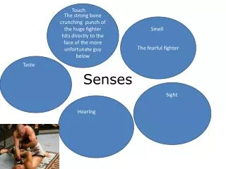
SENSES AND EAR. SENSES. Three types of senses: 1. SOMATIC SENSES 2. PROPRIOCEPTORS 3. SPECIAL SENSES. SOMATIC SENSES. 1. SOMATIC SENSES : Light touch (being touched by a feather), heat, cold, vibration, pressure, pain .

E N D
SENSES • Three types of senses: • 1. SOMATIC SENSES • 2. PROPRIOCEPTORS • 3. SPECIAL SENSES
SOMATIC SENSES • 1. SOMATIC SENSES: Light touch (being touched by a feather), heat, cold, vibration, pressure, pain. • These are routinely tested by doctors in a physical exam, especially for people with diabetes and lupus.
SENSES • 2. PROPRIOCEPTORS are found in the muscles, joints, and tendons. They measure the amount of movement, force, and position of the body. • Proprioception is often tested by having the patient close their eyes and saying if their fingers are up or down. • Proprioceptors send information to the cerebellum. That’s how you know your legs are crossed before you stand up. • Somatic senses (including pain) and proprioception are NOT considered special senses.
SENSES • 3. SPECIAL SENSES: Smell, taste, vision, hearing, equilibrium (balance).
OLFACTORY SENSE (smell) • Olfactory receptors are CHEMORECEPTORS; a special type of neuron which senses particular chemicals and triggers an action potential. • Chemoreceptors are at the roof of the nasal cavity. There are hundreds of thousands of types, and they can smell a wide variety of substances. • They are extremely sensitive, and can detect parts per billion, as in the scent of natural gas…just a few molecules! • The olfactory nerve goes through the cribiform plate to the OLFACTORY BULB (one of the shortest nerves in the body) and into the limbic system.
OLFACTORY SENSE (smell) • Scientists who are trying to find a way to make neurons divide to heal nerve injuries often study the body’s only mitotic neurons (undergo mitosis). • These neurons are the olfactory receptors. • People who experience imaginary odors have what are called “unicate fits”.
Olfactory Receptors Figure 16.3a, b
GUSTATORY SENSE (taste) • Sensed on taste buds, which are located mostly on the tongue surface, but are also on the palate, pharynx, and a few on the lips. • Taste buds have specialized cells, which increase surface area and have chemoreceptors. • They are surrounded by support cells (like glia). They synapse on sensory neurons, which go to the facial nerve. • Someone with a damaged facial nerve can not easily taste sweet, sour, or salty substances. Taste buds are the only parts of the nervous system that can regenerate completely. • The taste information is sent to the primary gustatory (taste) cortex, located in the parietal lobe of the brain.
Taste Buds Figure 16.1a, b
GUSTATORY SENSE (taste) • How many different tastes are there? Dozens. Salt, sweet, bitter, and sour are only a few. • Where are they located on the tongue? All tastes are located all over the tongue. • The picture in the book was drawn 120 years ago by an anatomist that knew his drawing was not right; he just wanted to use it as a starting point for further experimentation.
GUSTATORY SENSE (taste) • Taste appreciation is also involved in texture (a mealy apple is not as good), temperature (cold pizza tastes different than warm), and smell (perfume or cigarette smoke clog the senses and decrease taste). • There are dozens of taste receptors, hundreds of thousands of smell receptors, so the subtly of taste is from smell. • Foods people like are in opposite proportion to the numbers of taste receptors for that. People that love sweets have FEWER taste receptors for sweets, so they crave more taste of sweet things. If you dislike something, it’s because you have lots of receptors for it. Also, as you get older, you become less tolerant of sweets and more tolerant of bitter tastes (like beer and coffee).
Fun Facts • The catfish has over 27,000 taste buds. (What could be so tasty on the bottom of a pond?) • Flies taste with their feet.
THE EAR • Outer Ear • Middle Ear • Inner Ear
OUTER EAR • 1. OUTER EAR consists of the PINNA and the EXTERNAL AUDITORY CANAL. • The pinna is the cartilage of the ear; it acts as a funnel to capture the sound. • If you cup your hands to your ears (do it now), you’ll notice the sound of my voice is louder. • If you rolled up a piece of paper like a funnel and put it to your ear, it functions like the pinna. • The transmission of sound vibrations through the outer ear occurs chiefly through AIR.
The Outer (External) Ear Figure 16.17a
Ear Crease and Heart Disease • A diagonal earlobe crease is a potential indicator of coronary artery disease. This crease is called “Frank’s sign” • Whereas a “normal” earlobe is smooth, an earlobe with a crease has a fold, straight line, or wrinkle that appears to cut the earlobe in half. • Having an ear crease predicts an 80% chance of coronary artery disease in individuals younger than 40 years. The poor vascularity of this area allows it to show signs of clogged arteries.
MIDDLE EAR • 2. MIDDLE EAR is an AIR filled space with structures. • The TYMPANIC MEMBRANE (ear drum) vibrates in response to sound. • Attached to it are 3 bones: The MALLEUS (hammer), INCUS (anvil), and the STAPES (stirrup) are the smallest bones in the body. Together, they are only one inch long. • Their function is to amplify sound vibrations. The malleus vibrates the incus, which vibrates the stapes.
Structures of the Middle Ear Figure 16.17b
MIDDLE EAR • The middle ear is open to the nasopharynx by way of the AUDITORY TUBE (also called eustachian tube or nasopharyngeal tube), which is only the thickness of a pencil lead. • If this tube is closed, the ears feel plugged up. • The function of the auditory tube is to equalize the pressure of the middle ear and the outside air so the ear bones can vibrate. • Tubes are put in the tympanic membrane to drain fluids in kids with frequent ear infections.
Ear Tubes Doctors issue new guidelines for treating kids' ear infections, http://fxn.ws/YRnUv3
Structures of the Middle Ear Figure 16.17b
INNER EAR • 3. INNER EAR exists within the temporal bone (petrous portion). • It is a complex structure. It is located in a bony cavity called the BONY LABYRINTH (“maze”). • The bony labyrinth is filled with a fluid called PERILYMPH, which is similar to CSF. The bony labyrinth is the only place where perilymph is found.
The Inner (Internal) Ear Figure 16.17b
Inner Ear • Within the bony labyrinth is a snail-shaped structure, called the MEMBRANOUS LABYRINTH, which is filled with ENDOLYMPH. • The snail-shaped structure is divided into two main components. One is the COCHLEA (“snail shell”). This is responsible for hearing. • The other structure is responsible for balance and consists of three parts: • Semicircular Canals • Utricle • Saccule
Semicircular canals: Superior Posterior Lateral Utricle Saccule Vestibulocochlear nerve Cochlea Stapes
Inner Ear: Cochlea • Inside the cochlea are special neurons called HAIR CELLS; their axons form CN VIII. • The stapes is attached to the OVAL WINDOW, and vibrations cause the endolymph to vibrate; the hair cells here transmit this vibration. • Therefore the HAIR CELLS in this region are receptors for HEARING.
Organ of Corti • The sensory organ in the cochlea, which sits along the entire length of the basilar membrane and contains the sensory hair cells.
Within the organ of Corti, there is a difference in electrical charge (electrical potential) between inside and outside the hair cells.
Organ of Corti • When the basilar membrane moves in response to sound waves entering the cochlea, the organ of Corti moves with it, and this movement causes inner hair cells in a small area to open tiny transport gates (similar to trapdoors), which allows electrically charged ions to flow through. This causes an action potential to be sent along the auditory nerve to the brain. • The area from which the action potential arises provides information to the brain as to the pitch of the sound (rather as if the organ of Corti was a piano), and the rate of nerve firing provides information about the loudness of the sound. ORGAN OF CORTI VIDEO • HOW HEARING WORKS VIDEO
Cochlea • Low frequencies (like the longer strings of a piano) cause a response in the tip of the cochlea. • High frequencies cause a response at the larger end of the cochlea.
Cochlea • The axons of the hair cells form CN VIII, the VESTIBULOCOCHLEAR NERVE, which takes the signals to the brain. • Therefore, the cochlea is where the hearing receptors are located, so the cochlea is responsible for all of the hearing of sounds. • However, the ear does more than just hear; it is also responsible for balance and equilibrium.
VESTIBULAR SYSTEM • This system regulates balance. • It is also within the inner ear. • SEMI-CIRCULAR CANALS (Three of them, all in different planes) determine your head’s movement in three planes. • Within each semi-circular canal is endolymph and hair cells, whose axons go to the cerebellum.
When you move in one direction, like sliding across the room, the fluid sloshes like a cup of coffee, and it triggers the hair cells.
Utricle and Saccule • Attached to the semi-circular canals are two joined structures called the UTRICLE and the SACCULE. • These also contain HAIR CELLS and ENDOLYMPH. • Within the endolymph here are OTOLITHS (“ear rocks”) which are calcium deposits. • When you stand perfectly upright, these otoliths float above the hair cells, but if you tip your head to the side, they will land on and stimulate the hairs on that side. • That tells you what position your head is in and gives you a sense of equilibrium. • Therefore, the HAIR CELLS in this region are receptors for equilibrium and the OTOLITHS are an essential component of this process.
Anatomy and Function of the Otoliths Figure 16.21b
Ear Problems • Inflammation of the semi-circular canals give you a sense of motion when you’re not moving. The symptom is called VERTIGO (dizziness). The diagnosis is LABYRINTHITIS. • This can be debilitating. • Sometimes only one canal is affected, so you only get dizzy if you turn your head one way. • It can be caused by sinus infections, excess salt consumption, viral infections, stress. It is more common in smokers than non-smoking.
Treatment: • Benign paroxysmal positional vertigo (BPPV) • The Epley maneuver helps relieve the dizziness and spinning symptoms by moving the calcium particles out of the sensitive semicircular canals and into another inner chamber of the ear, where they don’t cause symptoms. • The exercise simply involves lying down and rolling your head in such way so that the particles fall out. • Epley Maneuver 1http://www.youtube.com/watch?v=ZqokxZRbJfw • Epley Maneuver 2 http://www.youtube.com/watch?v=pa6t-Bpg494
Treatment: House Ear Clinic • House Ear Clinic • Dr. Luxford • Chapman Ave, Orange • ARTIFICIAL EARS VIDEO
Visual Vertigo • When your eyes get one set of information that conflicts with the vestibular structures, such as when you are high up in the air or strobe lights flashing, or reading in a car. • Whether the vertigo is from visual or vestibular disturbances, your body interprets the signals as a poison invasion, so it initiates a vomit reflex.
“Cauliflower Ear”Hematoma auris or Traumatic auricular hematoma Common in boxers and wrestlers A blood clot or other fluid collects under the perichondrium. This separates the cartilage from the overlying perichondrium that is its source of nutrients, causing the cartilage to die. This leads to a formation of fibrous tissue in the overlying skin.