Download
1 / 20
240 likes | 767 Views
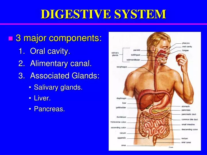
DIGESTIVE SYSTEM. 3 major components: Oral cavity. Alimentary canal. Associated Glands: Salivary glands. Liver. Pancreas. Wall of the Alimentary Canal. Four concentric layers: Mucosa : Epithelial Lining: Simple columnar epithelium (in stomach & intestine). Lamina propria :

E N D
DIGESTIVE SYSTEM • 3 major components: • Oral cavity. • Alimentary canal. • Associated Glands: • Salivary glands. • Liver. • Pancreas.
Wall of the Alimentary Canal Four concentric layers: • Mucosa: • Epithelial Lining: • Simple columnar epithelium (in stomach & intestine). • Lamina propria: • Connective tissue. • It contains mucosal glands. • Muscularismucosae: • 2 layers of smooth muscle cells. Serosa
Wall of the Alimentary Canal • Submucosa: Connective tissue containing blood vessels & nerves. May contain glands (as in duodenum). • MuscularisExterna: 2 smooth muscle layers: • Inner circular layer. • Outer longitudinal layer. • Serosa: Connective tissue covered by mesothelium (simple squamous epithelium). Serosa
STOMACH • It has 4 regions: cardia, fundus, body and pylorus. fundus cardia body pylorus
Fundus of Stomach • Mucosa: Shows gastric pitsin which open the fundic glands. • Epithelial lining:simple columnar epithelium (mucus-secreting).NO goblet cells. • Lamina propria: Connective tissue containing numerous fundic glands. • Muscularismucosae: 2 layers of smooth muscle.
Fundus of Stomach • Submucosa: • Connective tissue containing blood vessels & nerves. • NO glands. • MuscularisExterna: • 3 smooth muscle layers: • Inner oblique. • Middle circular. • Outer longitudinal. • Serosa: • Connective tissue covered by mesothelium.
Fundic Glands • Simple branched tubular glands. • Have short narrow ducts (pits). • Numerous; occupying most of the lamina propria. • Perpendicular to the surface.
Fundic Glands Composed of 5 cell types: • Parietal cells: secrete HCl and gastric intrinsic factor that helps absorption of vitamin B12. • Peptic (chief) cells:secrete pepsin. • Mucous neck cells:secrete mucus. • Enteroendocrine cells:secrete hormones. • Stem cells: regenerative cells.
SMALL INTESTINE • It has 3 regions: duodenum, jejunum and ileum. • To increase surface area the mucosa has: • Villi. • Crypts (intestinal glands).
Duodenum • Mucosa: Shows villiand crypts. • Epithelium:simple columnar epithelium with goblet cells. • Lamina propria: Connective tissue containing intestinal glands (crypts). • Muscularismucosae: 2 layers of smooth muscle cells.
Duodenum • Submucosa: • Connective tissue containing blood vessels and nerves. • Contains Brunner’s glands (secrete mucus). • MuscularisExterna: • 2 smooth muscle layers: • Inner circular layer. • Outer longitudinal layer. • Serosa: • Connective tissue covered by mesothelium.
Intestinal Glands (Crypts) • Simple tubular glands that open between villi. • Composed of 5 cell types: • Columnar cells: absorptive. • Goblet cells: secrete mucus. • Paneth cells: secrete lysozyme (antibacterial). • Enteroendocrine cells: secrete hormones. • Stem cells: regenerative cells.
LARGE INTESTINE • It is composed of: • Appendix, • Cecum, • Colon (ascending, transverse, descending & sigmoid), • Rectum, and • Anal canal.
Colon • Mucosa: Shows onlycrypts(NO villi) • Epithelium:simple columnar epithelium with numerous goblet cells. • Lamina propria: Connective tissue containing numerous crypts.Cells of the crypts are the same as in small intestine but NO Paneth cells.Lymphatic nodules:frequent. • Muscularismucosae:2 layers of smooth muscle.
Colon • Submucosa: • NO glands. • MuscularisExterna: • Inner circular & outer longitudinal smooth muscle layers. • The longitudinal layer is not continuous but in the form of 3 ribbons (teniae coli). • Serosa: • C.T. covered by mesothelium. • Has fat-filled pouches called appendices epiploicae.
Fundusof Stomach Colon Duodenum