Download
1 / 33

E N D
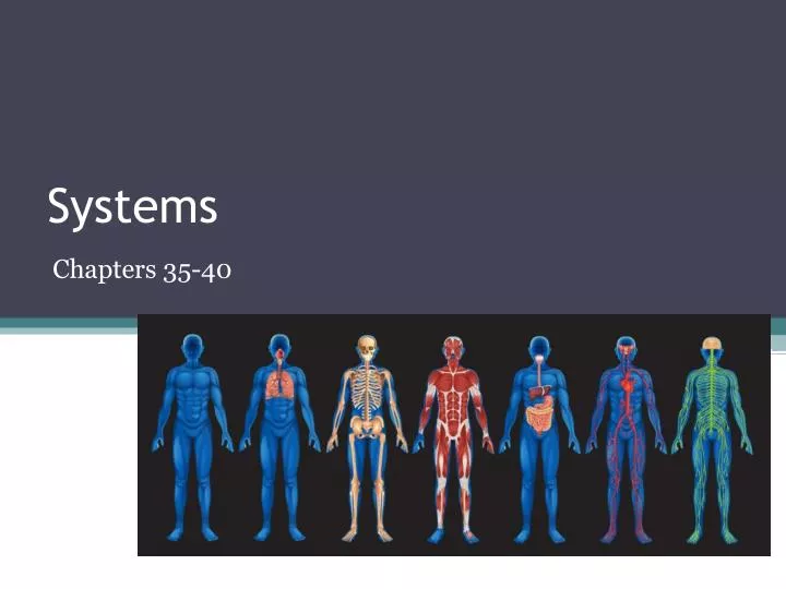
Systems Chapters 35-40
Chapter 35: The Nervous System • Function: Controls and coordinates functions throughout the body and responds to internal and external stimuli • Structures: • Neurons • Brain • Spinal Cord
Nervous System • Neurons – cells that transmit electrical signals called impulses • Structure of a neuron: • Cell body—contains the nucleus and much of the cytoplasm. • Dendrites—short, branched extensions that spread out from the cell body. • Axon—long fiber that carries impulses away from the cell body. • Myelin sheath—insulating membrane that surrounds the axon
Nervous System • Brain – Contains approximately 100 billion neurons and has a mass of about 1.4 kg. • Structures: • The cerebrum—responsible for the voluntary, or conscious, activities of the body. • The cerebellum—located at the back of the skull and coordinates and balances the actions of muscles. • The brain stem—connects the brain and spinal cord. • Two regions: pons and the medulla oblongata • Controls blood pressure, heart rate, breathing and swallowing • Thalamus and hypothalamus • Thalamus—receives messages from all of the sensory receptors throughout the body and then relays it to the proper region of the cerebrum. • Hypothalamus—control center for hunger, thirst, fatigue, anger and body temperature.
Nervous System • Spinal Cord • Main communications link between the brain and the rest of the body. • Thirty-one pairs of spinal nerves branch out from the spinal cord, connecting the brain to all the different parts of the body. • Reflex—quick, automatic response to a stimulus. • Allows the body to respond immediately
Nervous System • Sensory Receptors (5 types) • Pain Receptors –respond to chemicals released by damaged cells • Thermoreceptors – detect variations in temperature (skin, body core, & hypothalamus) • Mechanoreceptors – sensitive to touch, pressure, stretching of muscles, sound and motion • Chemoreceptors –sensitive to chemicals in the external environment (nose and taste buds) • Photoreceptors – sensitive to light (eyes) • Video
Chapter 36: Skeletal System • Function: supports the body, protects internal organs, provides for movement, stores mineral reserves, and provides a site for blood cell formation • Structures: • Bones • Ligaments
Chapter 36: Skeletal System • Bones • A solid network of living cells and protein fibers that are surrounded by deposits of calcium salts. • Periosteum—is a tough layer of connective tissue surrounding the bone. • Haversion canals—network of tubes running through compact bone containing blood vessels. • Bone marrow—red and yellow • Red—produces red blood cells, some kinds of white blood cells and platelets • Yellow—is made up primarily from fat cells
Chapter 36: Skeletal System • Ligaments – tough connective tissue that hold bones together • Joint – place where one bone attaches to another bone • Immovable joints—allow no movement. Bone are interlocked and held together • Bones in the skull • Slightly moveable joints—small amount of restricted movement. • Between adjacent vertebrae • Freely moveable joints—movement in one or more directions • Ball-and-socket, hinge joints, pivot joints and saddle joints
Chapter 36: Muscular System • Function: Movement, however, the type of movement is dependent on the location and type of muscle present • Structures: • Skeletal Muscles • Smooth Muscles • Cardiac Muscles
Chapter 36: Muscular System • Skeletal Muscles—usually attached to bones. • Are large, striated, have many nuclei, and vary in length from 1 mm to about 30 cm. • Often called muscle fibers. • Responsible for voluntary movements.
Chapter 36: Muscular System • Smooth muscles—Usually not under voluntary control. • Spindle-shaped, has one nucleus, and is not striated. • Found the walls of hollow structures such as the stomach, blood vessels, and intestines. • Move food through your digestive tract, controls the way blood flows through your circulatory system, and decrease the size of your pupils in bright light. • Can function without nervous stimulation.
Chapter 36: Muscular System • Found in just one place in the body—the heart. • Is striated like skeletal muscle—cells are smaller. • Usually have one nucleus, but may have two. • Not under the direct control of the CNS and are connected to their neighbors by gap junctions.
Chapter 36: Muscular System • How muscles and bones interact • Skeletal muscles are joined to bones by tough connective tissues called tendons. • Most skeletal muscles work in opposing pairs. • When one muscle contracts the other relaxes. • Muscles of the upper arm. • A controlled movement requires contraction by both muscles.
Chapter 36: Integumentary System • Function: Serves as a barrier against infection and injury, helps to regulate body temperature, removes waste products from the body, and provides protection against ultraviolet radiation from the sun. • Structures: • Skin • Hair • Nails
Chapter 36: Integumentary System • Skin: two layers • Epidermis • Dermis • Epidermis—outer layer of the skin • The outside of the epidermis is made up of dead cells. • Inner layer is made up of living cells. • Dermis—inner layer of skin • Interacts with other body systems to maintain homeostasis by helping to regulate body temperature.
Chapter 36: Integumentary System • Hair and Nails • Basic structure is keratin • Hair • Protects the scalp from ultraviolet light. • Provides insulation from the cold. • Prevent dirt and other particles from entering the body. • Nails • Grow at an average rate of 3 mm per month. • Grow from an area of rapidly dividing cells. known as the nail root.
Chapter 37: Circulatory System • Functions: Transportation system of the body and is involved in respiration, nutrition, waste removal, immunity, and thermal regulation • Structures: • Heart • Vessels • Blood
Chapter 37: Circulatory System • The Heartis a hollow organ that is about the size of your clenched fist. • Contracts on average 72 time a minute, pumping about 70 mL of blood with each contraction. • Each side contains two chambers— the upper chamber, which receives blood is the atrium, and the lower chamber, which pumps blood out of the heart, is the ventricle. • The Heart
Chapter 37: Circulatory System • Circulation through the heart • Blood enters the right atrium of the heart, from the rest of the body, through the superior or inferior Vena Cava. • From the right atrium it moves to the right ventricle where it is pumped to the lungs through the pulmonary arteries. • The now oxygen-rich blood returns to the heart through the pulmonary veins and enters the left atrium. • Lastly, the blood moves down into the left ventricle were it is pumped to the rest of the body through the aorta.
Chapter 37: Circulatory System • Vessels • 3 types • Arteries—carry blood away from the heart • Main artery leading away from the heart is the Aorta • Capillaries—have one layer of cells where diffusion and exchange of materials takes place • Veins—carry blood back to the heart • Blood reenters the heart through the inferior and superior Vena Cava
Chapter 37: Circulatory System • Blood • Red Blood Cells—transport oxygen • Hemoglobin—iron-containing protein that binds to oxygen in the lungs and transports it. • White Blood Cells—guard against infection, fight parasites, and attack bacteria. • Platelets—plasma proteins that make blood clotting possible • Hemophilia—genetic disorder in the blood clotting pathway.
Chapter 37: Respiratory System • Function: Basic function is to bring about the exchange of oxygen and carbon dioxide between the blood, the air, and tissues. • Structures: There are 3 major parts of the respiratory system: • The airway • Lungs • Muscles of respiration.
Chapter 37: Respiratory System • The airway • The airway includes the nose, mouth, pharynx, larynx, trachea, bronchi, and bronchioles • Pharynx—serves as a passageway for both air and food. • Larynx—contains two highly elastic folds of tissue known as the vocal cords. • Trachea—windpipe • Bronchi—two large passageways that lead from the trachea to the lungs.
Chapter 37: Respiratory System • The lungsare a pair of spongy, air-filled organs located on either side of the chest • There are 150 million alveoli in each healthy lung. • Oxygen dissolves in the moisture on the inner surface of the alveoli and then diffuses across the thin-walled capillaries into the blood. • Carbon dioxide diffuses in the opposite direction.
Chapter 37: Respiratory System • Muscles of Respiration • Diaphragm—large, flat muscle located at the bottom of the chest cavity. • When you breathe in, or inhale, the diaphragm contracts and the rib cage rises up. • Creates a partial vacuum. • Atmospheric pressure then fills the lungs with air. • Exhaling is passive. The rib cage lowers and the diaphragm muscle relaxes and the pressure in the chest cavity becomes greater than atmospheric pressure
Chapter 38: Digestive System • Function: The digestive system is a group of organs working together to convert food into energy and basic nutrients to feed the entire body • Structures: • Mouth, pharynx, esophagus, stomach, liver, small intestine, and large intestine
Chapter 38: Digestive System • Mouth • Chewing begins the process of mechanical digestion—the physical breakdown of large pieces into smaller pieces. • Saliva—contains amylase, an enzyme that breaks the chemical bonds in starches and releases sugars. • Esophagus • Food tube—carries food from the mouth to the stomach
Chapter 38: Digestive System • Stomach—large muscular sac that continues the mechanical and chemical digestion of food. • Chemical digestion—pepsin (an enzyme) and hydrochloric acid begins the process of protein digestion • Mechanical digestion—muscles contract to churn and mix stomach fluids and food producing a mixture known as chyme. • Liver—produces bile which assists in breaking down fats
Chapter 38: Digestive System • Small intestine—location of most of the chemical digestion and absorption of the food you eat • The folded surface of the small intestine is covered with fingerlike projections called villi. • Each villi is covered by thousands of fingerlike projections called microvilli. • Provide and enormous surface area for absorption. • Large intestine—function is to remove water from the undigested material that is left.
Chapter 38: Excretory System • Function: The excretory system maintains the homeostasis of several important internal conditions by controlling the excretion of substances out of the body. • Structures: • Kidneys • Bladder
Chapter 38: Excretory System • Kidneys—remove waste products from the blood; maintain blood pH; and regulate the water content of the blood, and therefore, blood volume. • Located on either side of the spinal column near the lower back. • Activity of kidneys is controlled by the composition of blood itself. • Bladder—saclike organ where urine is stored before being excreted
Chapter 38: Excretory System • If anything goes wrong with the kidneys, serious medical problems follow. • Transplant of a healthy kidney from a compatible donor. • Kidney dialysis—blood is removed from the body through a tube and pumped through special tubing that removes waste products. • The purified blood is then returned to the body.