Download
1 / 32
370 likes | 449 Views
Explore the clinical applications and functionality of pelvic floor imaging using ultrasound and MRI techniques, crucial for assessing pelvic muscle function and disorders such as incontinence, prolapse, and perineal pain.

E N D
Imaging of the pelvic floor:Ultrasound and MRI Dr Bruce AllenHorizon Radiology Acknowledgement to Dr Hans Dietz and Dr Jenny Kruger
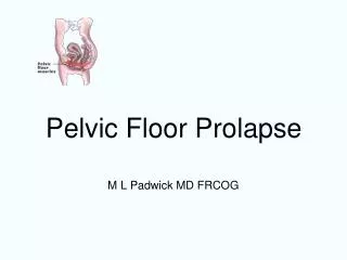
Clinical application of Pelvic floor imaging • Pelvic floor muscles involved in: • Maintenance of continence • Support of the organs of the pelvis • Vaginal delivery • Failure of these muscles increases risk of: • Urinary and fecal incontinence, • Prolapse of the organs of the pelvis, • Perineal pain and dyspareunia.
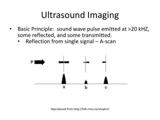
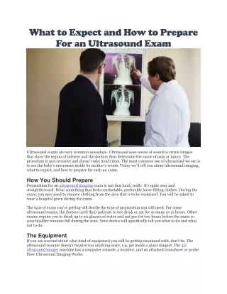
Ultrasound imaging • Cost effective • Do not need expensive machines to do basic imaging • Ultrasound is highly operator dependant • Not difficult to learn • Real time, functional studies easy • MRI • Expensive • Images are easier to understand • Functional studies difficult.
2D imaging - Ultrasound • Until recently 2D ultrasound scanning only methodology used define pathology and normal function of pelvic floor • Abdominally, transvaginal or translabial • Descent of bladder neck, uterus and rectal ampulla during a valsalva • Transperineal ultrasound useful biofeedback measure for patients • Image is in mid-sagittal plane • $12,000 machine.
Translabial 2D Ultrasound • Patient is supine, bladder empty ( or standardized filling) • Knees flexed, feet on the table • Transducer covered in glove/condom for hygiene • Placed fairly firmly on the perineum in the mid sagittal orientation.
Typical 2D image of the pelvic floor muscles urethra cranial
MRI: Sag midline, normal anatomy • Bony landmarks • 20 mins scan time • Anatomy
Pelvic floor functional assessment • Training • Contraction • Valsalva
Pelvic floor muscle contraction Contraction assess: 1. Narrowing of the hiatus in the AP diameter 2. Movement of the bladder neck 3. Strength of the PF muscle
Effective valsalva manouevre • Valsalva assess: • Descent of bladder, uterus, rectum. Urethral rotation. • Development of cystocele, prolapse or rectocele • Width of hiatus in the AP diameter
MRI: Valsalva. Cystocoele • Functional: • 4 min per sequence • Valsalva • defaecation • (training)
2D imaging • Measurements of bladder neck descent and urethral rotation. 2D Imaging Ultrasound images showing measurement of bladder neck descent and urethral rotation. Bladder neck descent (BND)= x-r –x-s. (Dietz et al 2004)
Clinical use of 2D ultrasound • Still widely used • Bladder, uterine and rectal descent. • Bo, K. and M. Sherburn, Evaluation of female pelvic-floor muscle function and strength. Physical therapy, 2005. 85(3): p. 269-82, Mar. • Abdominal ultrasound • Athanasiou, S., et al., Direct imaging of the pelvic floor muscles using two-dimensional ultrasound: a comparison of women with urogenital prolapse versus controls.BJOG: An International Journal of Obstetrics and Gynaecology, 2007. 114(7): p. 882-888. • Endovaginal probe • Costantini, S., et al., Perineal ultrasound evaluation of the urethrovesical junction angle and urethral mobility in nulliparous women and women following vaginal delivery. Int Urogynecol J Pelvic Floor Dysfunct, 2005. 16(6): p. 455-9. • Transperineal ultrasound • Dietz, H., Pelvic Floor Ultrasound. Current Medical Imaging Reviews, 2006. 2: p. 271-290. • Dietz, H., B. Haylen, and J. Broome, Ultrasound in the quantification of female pelvic organ prolapse. Ultrasound in Obstetrics and Gynecology, 2001. 18: p. 511-514.
3D ultrasound imaging • 3D ultrasound widely used in obstetric scanning so equipment is now readily available • $100,000 – $250,000 • Acquisition of volume images allow access to the ‘axial’ plane – previously domain of magnetic resonance imaging.
Protocol for 3D pelvic floor imaging • Translabial imaging: • Imaged supine after voiding • Transducer ‘sits’ on the perineum mid-sagittal orientation • Mid-sagittal image on the screen • Symphysis pubis reference point – during movement • Methods highly reproducible (Guaderrama, Yang, Dietz ).
3D US pelvic floor imaging – levator hiatus Voluson 730 expert system. (Dietz et al 2005)
3D pelvic floor ultrasound – assessing function • Levator hiatus: • ‘plane of minimal dimensions’ • Smallest distance from the inferior edge of the symphysis pubis to the anal rectal angle • Levator hiatal area bounded by the symphysis pubis anteriorly, anal rectal angle posteriorly, puborectalis/ pubococcygeus laterally. • Hiatal area measures pelvic floor function • Rest • Maximum pelvic floor muscle contraction • Maximum valsalva • (Training).
3D imaging: hiatal measurement A mid-sagittal image. Line indicates plane of minimal dimensions B corresponding ‘axial’image showing entire levator hiatus
Conclusions • Translabial ultrasound 2D / 3D /4D • Function and anatomy • effective, easy, low cost method for assessment of the PF • Used to confirm/or not the digital diagnosis of PF dysfunction • Biofeedback training • MRI • Anatomy (and function)