Download
1 / 38
440 likes | 1.15k Views
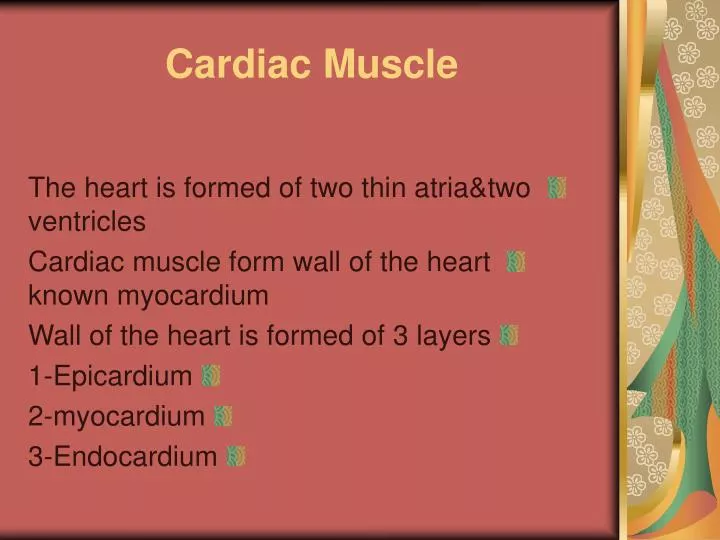
Cardiac Muscle. The heart is formed of two thin atria&two ventricles Cardiac muscle form wall of the heart known myocardium Wall of the heart is formed of 3 layers 1-Epicardium 2-myocardium 3-Endocardium. Epicardium:

E N D
Cardiac Muscle • The heart is formed of two thin atria&two ventricles • Cardiac muscle form wall of the heart known myocardium • Wall of the heart is formed of 3 layers • 1-Epicardium • 2-myocardium • 3-Endocardium
Epicardium: • Visceral layer of pericardium formed of simple sq.epithelium&layer of connective tissue • Myocardium: • formed of cardiac muscle
Endocardium: • lines the heart from inside • Having four layers from inside • 1-simple sq.endothelium • 2-subendothelial layer of dense fibrous c.T. • 3-dense elastic&collagenous membrane • 4-loose of cT.layer contain BV.&purkinje muscle fibre
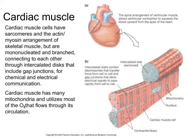
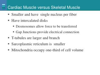
Characteristic of C.Muscle • -individual muscle cells surround by cT endomysium contain Bv.&lymphocytic • - small diameter • -branch&join each other forming sheets • -contract spontaneous(involuntaryin action) • -striation: have irregular striation • -nuclei are large,central in postion&oval in shape
-cytoplasm granular acidophilic sarcoplasm rich in glycogen,mitochondria&lipochrome granules Diad System is formed of transverse T-Tubules are present at the level of Z-Lines
The cardiac muscle transversed by Dark staining discs which extend across the fibres called Interecalated Discs
The Intercalated Disc • There are 3 types of junctional complex at the intercalated discs • 1-Desmosomal type of junction which prevent sepration of the muscle cells • 2-Adherens type of junction • 3-Gap type of junction impulses between heart muscles • The atrial muscle fibres secret Atrial naturetic Diuretic Hormone(ANDH)
Cardiac muscle have the ability to undergo rhythmic contraction • Valves of the heart are formed of dense CT. rich in elastic &collagenous fibres and covered by simple sq. endothelium,
Phagocytic histiocyte cells are present in the CT.of the valves in order to englulf any micro-organism • Fibrous skeleton of the heart formed of dense fibrous tissue • Cardiac muscle&valves are attached to the fibrous skeleton
INTERCALATED DISCS Intercalated Discs Cardiac Muscle Fibers (transverse section)
INTERCALATED DISCS Gap Junctions Desmosomes
The conducting system of the heart • Its avascular neuro-muscular structures consist of • 1-sino-atrial node(SA) in the Rt.atrium,its pacemaker of the heart • 2-Atrio-ventricular(AV) node present in septal wall of Rt.atrium • 3-atrio-ventricular bundles of His:branches into Rt.&Lf bundles • 4-Rt&Lf. Bundles branches:the Rt. Branch is called moderator Band
The moderator Band • Formed of: • Cardiac muscle bundles,fat cells,Blood capillaries,Purkinje muscle fibres
Purkinje Muscle Fibres • composed of elongated cells called Purkinje Cardiac Muscle • they are larger in diameter than the cardiac fibres • they are paler in color • they are usually grouped into bundles • each purkinje fibre is formed of separated,short,thick,elongated cylindrical cells
The sarcolemma of purkinje fibres is thin and irregular • cytoplasm is granular and rich in glycogen has myofibrils which are peripherally situated • Purkinje fibres have no diad tubular systems • The intracalated discs are few in purkinje muscle fibres
PURKINJE FIBERS Purkinje Fibers (specialized conducting tissue) PF Purkinje Fibers (trans. section) glycogen stores adipocyte
Nervous Tissue 1-Central Nervous System (CNS), consisting of the brain and spinal cord. 2-Peripheral Nervous System (PNS), consisting of nerve fibers, aggregates of nerve cells and glia and ganglia. • It is estimated that the human nervous system consists of at least 10 billion neurons. • Nervous tissue consists of two groups of cell types: • 1-Nerve cells (Neurons) • 2-Neuroglia.
Neurons • Neurons are post-mitotic structures that shortly after birth lose the ability to divide. • Further changes involve only reduced number of neurons (neuronal death), or changes in volume or in neuronal connections. • Neurons have two special properties: 1-Irritability (the ability to respond to a stimulus) 2-Propagation of impulses (the ability to conduct impulses).
The morphofunctional unit of the nervous system is the neuron. • Similar to the Cell Theory, which stipulates the cell as the basic building block of the body, the Neuron Theory describes the neuron as the basic building block of the nervous system, and that the nervous system functions through transmission of information through networks of neurons.
Most neurons have three main parts: 1-Dendrites 2-Perikarya (cell bodies) 3-Axon • The dendrites are receptive to stimuli and bring stimuli from the environment (sensory epithelial cells or other neurons) to the cell body. There are usually several dendrites per neuron. • The perikaryon (cell body) is also receptive to stimuli, but also serves as the trophic or synthesizing center for the whole nerve. • The axon is a long process emerging from the cell body. There is only a single axon for each neuron. The axon transmits impulses to other neurons, or to effectors: muscle or gland cells. The distal portion of the axon is usually branched (terminal arborization). • Neurons and their processes are very variable in form and size. Some neurons are very large (with perikarya of up to 150m), whereas others are very small (perikarya of only 4-5m).
ANATOMY OF A NEURON impulse conduction axons dendrites axo-somatic synapse Nissl’s substance axo-dendritic synapse boutons axon axon hillock axo-axonic synapse myelin sheath oligodendrocyte unmyelinated collateral myelin sheath central nervous system (CNS) myelin sheath peripheral nervous systen (PNS) Schwann cell motor end plate synaptic vesicles motor end plate
MOTOR NEURON CELL BODY (H&E) DENDRITIC SPINES (boutons)-silver axo-denritic synapses dendrite (boutons) Nissl’s substance nucleolus dendrite nucleus axon
Morphological classification of neurons • Neurons are classified according to the size, number and shape of their processes. 1-Unipolarneurons have a single process (axon). These are found in sensory ganglia of dorsal roots of spinal nerves. 2-Pseudo-unipolar neurons have two processes (one dendrite and one axon). These are very rare and have a limited distribution in the body. They are present in special sensory structures including the retina, olfactory epithelium, and vestibular and cochlear nerves). 3-Bipolar neurons possess several processes (several dendrites and a single axon). Most neurons belong to this category.
4-Multipolar nerve cells: • They are subdivid into 3 Subtypes • A-Stellate-shaped or polygonal cells • Present in anterior horn cells of the spinal cord&in simpathetic ganglia • B-pyramidal cells: present in cerebral cortex • C-Pyriform cells flask in shape cells as purkinje cells of the cerebellum
NEURON TYPES Bipolar Neuron (retina & olfactory epithelium) Pseudounipolar Neuron (sensory spinal reflex arc) Unipolar Neuron (embryonic)
Physiological classification of neurons • Neurons may also be classified according to their function. 1-Sensory neurons. These receive sensory stimuli from the environment (from receptors) and from within the body (e.g. unipolar neurons). 2-Motor neurons. These control the effector organs (muscles, exocrine glands, endocrine glands)
3-Interneurons (Intermediate neurons). These are typically found in the CNS and connect between other neurons (often between sensory and motor neurons). 4-Neurosecretory neurons. These are specialized neurons that synthesize and secrete hormones.
PERIPHERAL NERVE ENDINGS-(MUSCLE) visceral visceral sensory (proprioreceptors) motor motor unit Neuromuscular Spindle motor end plates (proprioreceptors (boutons)
Each neuron has 3 physiological parts or segments: 1-Receptive segment (dendrites and perikaryon). The perikaryon also has an additional trophic and synthesizing role. 2-Conductive segment(axon) 3-Transmissive segment(synapse).
Differences Between axon and dendrites • Axon: • 1-single process • 2-thin&long • 3-uniform diameter along its length • 4-branches&also give collateral • 5-contains neurofibrils but no nissl granules • 6-Two types of axonal transport exist Antigrade&retrograde
The Dendrites • 1-usually multiple process • 2-usually short&thick • 3-their thickness decreases gradually towards its end • 4-have many fine side projection called spines • 5-Contain Nissl granules&neurofibrils • 6-receive impulses from other neurones
Classifiction of Neurones • According to the length of their axons • -Golgi Type1Neuron;have long axons as the neurones of cerebral cortex • -Golgi type 2 Neuron: have short axons as neurones of retina