Download
1 / 38
400 likes | 580 Views
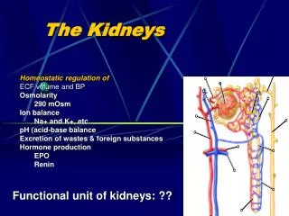
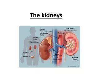
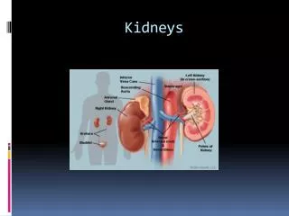
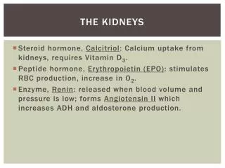
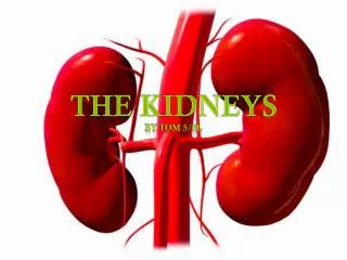
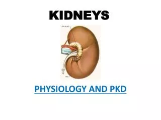
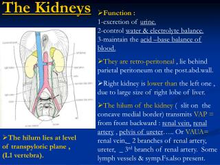
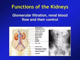
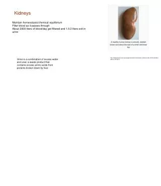
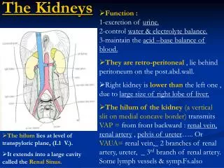
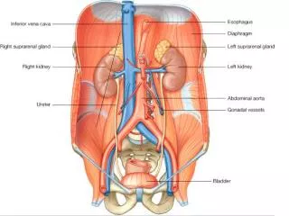
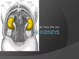
Regulation of extracellular components. Elimination of waste products. Regulation of acid-base balance. Erythropoietin production. Formation of renin. Xenobiotics metabolism. Production of glucose and 1,25-dioH vitamin D3. The Kidneys. Anatomy of kidney. Nephron.

E N D
Regulation of extracellular components. Elimination of waste products. Regulation of acid-base balance. Erythropoietin production. Formation of renin. Xenobiotics metabolism. Production of glucose and 1,25-dioH vitamin D3. The Kidneys
Heavy metals accumulate in body Induction of metallothionein by liver. Cadmium (Cd) stimulates its production. Metallothionein bind with Cd and protects other organs from its toxic effects. The kidney is vulnerable to this complex. Beta-2-microglubulin excretion from kidney is diagnostic of Cd toxicity. Nephrotoxicants
Long term use of Analgesics Antibiotics. Anticancer drugs. Ochratoxin A. Nephrotoxicants
Immunosupression by toxicant • Halogenated Aromatic Hydrocarbons 2,3,7,8-tetrachlorodibenzo-p-dioxin(TCDD) Lymphoid atrophy, Thymic involution. Polychlorinated biphenyls (PCBs) Polybrominated biphenyls (PBBs) Suppress Antibody Response. • Formaldehyde & Isocyanates Types I and IV reactions
immunosupression by toxicant • Polyaromatic hydrocarbons (PAHs) Depress Humoralimmunity , Cell mediated immunity and Tumor resistance. • Nitrosamines (dimethyl & diethyl nitrosamine) inhibit T-cell dependent humoral immune response. • PesticidesImmunotoxic
SKIN • Physical protection from environmental agents • Hydroregulation through active &passive mechanism • Thermoregulation to maintain core body temperature. • Chemical synthesis of vitamin D. • Immunological surveillance and function. • Absorption of pharmaceutical preparation. • Sensory reception of pain, temperature, touch & pressure
Toxicological point of view • Route of exposure for systemic toxicants. • Direct target for toxicity. • Xenobiotic metabolizing organ. • Minor pathway for the elimination of certain toxicants.
Epidermis: outermost layer; mostly epithelial cells; non-vascular Dermis: fibrous connective tissue; vascular Hypodermis: (superficial fascia)not skin; protective; adipose and loose connective tissue Epidermis is thick keratinized Stratified Squamous Epithelium consisting of four cell types and five layer Keratinocytes Melanocytes Merkel cells Langerhan’s cells SKIN
. Stratum basale(stratum germinativum)—deepest layeri. Single layer of mitotically active cells ii. Give rise to keratinocytes (youngest) iii. Includes melanocytes and some Merkel cellsStratum spinosum(Prickly layer)—weblike network of cells formed by intermediate filaments attached to desmosomesi. Comprised of keratinocytes ii. Includes melanin granules and Langerhans cellsStratum granulosum—thick; 3-5 cell layers; keratinocytes are modifiedi. Flattened; nuclei and organelles lost ii. Keratohylaline and lamellated granules accumulate iii. Lamellated granules are glycoproteins, released into extracellular space, that reduce water loss iv. Cells more resistant to destruction
Stratum Lucidum—a few rows of clear, flattened, dead keratinocytes; layer occurs only in thick skini. Keratohyalin granules—gummy substance associated with keratin filaments ii. Cells aggregate in parallel arraysStratum Corneum(Horny layer)—outer most layer; most of epidermis thicknessi. 20-30 cell layers thick ii. Keratin, thickened plasma membranes and glycoproteins protect against abrasion and loss of water iii. Cornified or horny cells—remnants of cells from this layer
Langerhans cells ANTIGEN PRESENTATION.
Merkel’s cells TOUCH RECEPTOR
Skin absorption of chemicals Percutaneous absorption:systemic toxicity from skin exposure can occur only when chemical moves from the epidermis into dermis of the skin, which contain blood vessel, the movement is by passive diffusion. The major barrier is stratum corneum. The rate of penetration is largely related to the lipophilicity of the chemical. (Lipophilic substances, Hydrophilic substances)
Skin toxicity: Local effects Contact Dermatitis • Irritant contact dermatitis • Allergic contact dermatitis • Phtotoxic skin response.
Effects of chemical exposure on skin Urticarial reactions:Type I allergic reactions cause the release of histamines, IgE, and local inflammatory mediators. Cutaneousgranulomas:Inflammatory response to insoluble materials. Hair loss:due to exposure of thallium, cancer therapeutic agents, depilatories. Hypopigmentation: inhibition/destruction of melanocytes (phenolic preparation, hydroquinone). Hyperpigmentation: Heavy metals, acridines Color change:orange/yellow from picric acid, green from copper dust, black from osmium trioxide.