Download
1 / 89
1.21k likes | 2.84k Views
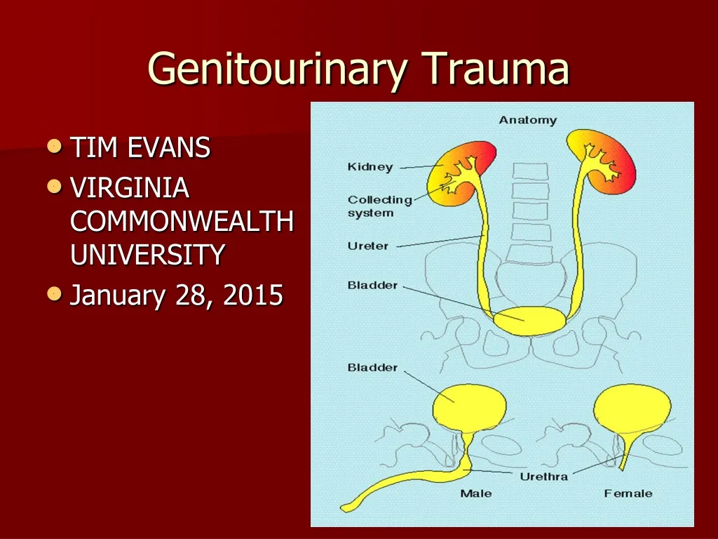
Genitourinary Trauma. TIM EVANS VIRGINIA COMMONWEALTH UNIVERSITY January 28, 2015. Background. If injury to GU system identified, multi-organ injury is the rule. Examples: If renal injury found following penetrating trauma, 80-95% chance of other significant injury

E N D
Genitourinary Trauma • TIM EVANS • VIRGINIA COMMONWEALTH UNIVERSITY • January 28, 2015
Background • If injury to GU system identified, multi-organ injury is the rule. • Examples: • If renal injury found following penetrating trauma, 80-95% chance of other significant injury • If renal injury found following blunt trauma, 75% chance of other significant injury found • Other injuries may be more immediately life threatening and therefore • GU injury diagnosis may be overlooked or delayed
Case • Patient #1 is a 25 year old male is struck in the flank with a baseball bat. His systolic blood pressure is always above 100 mm Hg and his exam is only remarkable for a flank hematoma without abdominal tenderness. His urinalysis shows no RBCs. • Patient #1 got pissed off at the guy who hit him so he shot Patient #2 in the flank. Patient #2 is hemodynamically stable and does not have any RBCs in his urine • Two Questions: • Do either of these reprobates need imaging? • Do we need more bat control legislation?
Renal Trauma • Most common GU injury—65% of GU injuries • 10% of abdominal injuries involve the kidneys • Mechanism • 80-95% due to blunt force—MVC, falls, assaults, sporting events
Renal Anatomy Retroperitoneal Adjacent to lower two thoracic and first four lumbar vertebrae Upper poles protected by ribs so lower poles more commonly injured Right kidney inferior to left and more commonly injured Kidney mobile, hilum more fixed—concern with shearing injury with deceleration
When are you concerned about renal injuries? • Mechanism of Injury • Penetrating injuries of abdomen, back or flank • Deceleration injuries • Physical exam • Tenderness of abdomen or flank • Ecchymosis of abdomen or flank • Xray • Fractures of lower ribs, thoraco-lumbar spine
When are you concerned about renal injuries? • Hematuria—over 95% of patients with renal trauma will have some degree of hematuria (>5 rbc/hpf) • THE PRESENCE OR DEGREE OF HEMATURIA DOES NOT CORRELATE WITH THE SEVERITY OF THE INJURY • 25% of patients with gross hematuria have minor injuries • 40% of the most serious renal injuries do not have any hematuria
Indications for imaging for renal trauma • Penetrating trauma in proximity to kidneys—the presence or absence of hematuria in penetrating trauma not predictive of injury, location of wound is most important factor • Gross hematuria • Microscopic hematuria (>3-5 RBC/HPF) with hemodynamic instability—systolic BP<90 at any time • Persistent microscopic hematuria • ?Significant deceleration mechanisms • ?Proximal injuries with blunt mechanisms Mee SL, et al: Radiographic Assessment of Renal Trauma: A ten-year prospective study of patient selection. J Urol 141:1095, 1989
When not to image in patients with concern for renal trauma • Patients with microscopic hematuria who have always been hemodynamically stable • Patients who are not hemodynamically stable
Microhematuria and no shock Gross hematuria or Microhematuria and shock (SBP<90 mmHg) all imaged-422 Imaged- Significant injury 3 Significant renal injuries 78 Imaged- Contusion 581 Without Imaging 1004 1 renal repair Renal repair 34 No significant renal injuries missed Miller KS, McAninch JW: Radiographic assessment of renal trauma. Our 15-year experience. J Urol 1995;154:352-355
Imaging techniques • Contrast enhanced CT—the best test, up to 98% accurate, not great for renal vein injuries • IVP—perhaps useful in the OR to determine function of contralateral kidney before contemplated nephrectomy • Angiography—better than CT for defining injuries to renal artery and vein, also used therapeutically to embolize or stent artery injury • Ultrasound—30% false negative rate for injury, used to look for two kidneys, free fluid • Contrast Enhanced Ultrasound—perhaps • MRI—not first line due to time, sensitivity similar to CT, can be used for follow up studies
AAST Kidney Injury Severity Scale—Revision 2011 • Grade IV - originally encompassed contained injuries to the main renal artery and vein, and collecting system injuries. Revision: adds segmental arterial and venous injury, and laceration to the renal pelvis or ureteropelvic junction. Multiple lacerations into the collecting system used to be considered a shattered kidney (Grade V), but now remains Grade IV. • Grade V - originally included main renal artery or vein laceration or avulsion, and multiple collecting system lacerations (shattered kidney). The revised classification includes only vascular injury (arterial or venous) and includes laceration, avulsion or thrombosis.
Trauma Penetrating Blunt Injury in proximity to kidney Hematuria (Gross or microscopic) Associated with shock (SBP <90) Hematuria Microscopic (>5 RBC/HPF) No shock Unstable Stable Image with concern for other organs Abdominal exploration CT scan with IV contrast Single-shot IVP on table Grades III-V Clinical follow-up Abnormal or inconclusive Selective renal exploration Renal exploration
Management of Renal Injuries • Grade I—home • Grade II-IV—admit, observe • Grade V—observe, vascular repair/stent, or nephrectomy Only absolute indications for surgery are persistent renal bleeding with hemodynamic instability, active extravasation of IV contrast, expanding or pulsatile perirenal hematoma suggesting Grade V vascular injury
Complications of Renal Injuries • Mortality 3% • Complications • First six weeks • Hemorrhage/shock • Sepsis/abscess • ATN • Late • Renovascular HTN 1-4%
CASE • 30 year old s/p cystoscopic removal of distal ureteral stone. Now with flank pain and nausea. T 39 C, diffuse abdominal and flank tenderness noted. • U/A--negative • Diagnosis? Studies?
Ureteral Trauma • Accounts for 1% of urologic trauma • Most commonly iatrogenic following GU, gynecologic, vascular or colorectal surgery • If following external trauma, 80-95% due to penetrating mechanism, usually GSW
Ureteral Anatomy • Thin, mobile tubes running between renal pelvis and posterior superior angle of bladder • Retroperitoneal in abdomen • Protected from injury by size and mobility
When are you concerned about ureteral injuries? • Recent GU, gynecologic, vascular or colorectal procedure • Penetrating (usually GSW) trauma to abd, back, flank • Deceleration mechanisms • Suspicion raised with injuries to iliac vessels, urinary bladder, sigmoid colon, thoracolumbar dislocations, lumbar spine (including process) fractures
Hematuria following ureteral injuries • Ureteral injury following iatrogenic cause—10-15% of patients with hematuria • Hematuria absent in 30-60% of identified ureteral injuries from external violence • Hematuria following penetrating trauma—a study of 71 ureteral injuries • 32% without hematuria • 40% with gross hematuria • 28% with microscopic hematuria Brandes SB, et al: Ureteral injuries from penetrating trauma, J Trauma 36:766, 1994.
IMAGING FOR URETERAL INJURIES • Most injuries diagnosed during laparotomy and no imaging ever done • Contrast CT with delayed imaging—most common findings are extravasation of contrast into medial perirenal space and absence of contrast in distal ureter if transected • Retrograde pyelogram • IVP—one shot IVP done in OR for penetrating trauma
Delayed CT images showing extravasation of urine from ureteral injury
Blunt trauma Penetrating trauma Gross hematuria, or microhematuria with deceleration or hypotension or associated injuries Stable, to CT + contrast + delayed films Unstable, to OR Gross or micro-hematuria Unstable, to OR Yes No Potential ureteral injury (ureteral nonopacification or extravasation) Abnormal Intraoperative One-shot IVP Normal Intraoperative one-shot IVP Normal Consider other sources for hematuria (bladder, urethra, kidney) Stent removal 6 weeks After stent removal consider periodic renogram or surveillance ultrasound (defect hydronephrosis to rule out recurrence Explore ureter and repair Bullet/knife wound in vicinity of ureter
American Association for the Surgery of Trauma (AAST) Ureter Injury Severity ScaleGradeDescription I Hematoma Contusion or hematoma without devascularization II Laceration <50% transection III Laceration >50% transection IV Laceration Complete transection with <2 cm devascularization V Laceration Avulsion with >2 cm devascularization
MANAGEMENT OF URETERAL INJURIES • Treatment • Stents—Grade 1 • Surgery—Grade 2 and above • Complications • Ureteral stricture • Fistula • Retroperitoneal fibrosis • Abscess/Sepsis
Intraoperative recognition Minor ureteral injury Major ureteral injury Stent Primary stented ureterourostony, psoas hitch, or flap with or without kidney mobilization Consider placement of percutaneous nephrostomy in rare case of extremely long injury Ureteral stent 6 weeks Follow-up retrograde pyelography and stent removal or replacement as needed Consider endoscopic methods (laser, balloon) Primary stented ureteroureterostomy After stent removal consider periodic renogram or surveillance ultrasound to rule our recurrence
Postoperative recognition CT with contrast (+ delayed films) + retrograde pyelography Minor ureteral injury Major ureteral injury Ureteral stent 6 weeks Attempted retrograde stent placement Follow-up retrograde pyelography and stent removal or replacement as needed Fail Success Success Percutaneous nephrostomy and anterograde stent placement, if possible Consider endoscopic methods (laser, balloon) Primary stented ureteroureterostomy, psoas hitch or flap Consider autotransplant or ileal loop in rare case of extremely long injury After stent removal consider renogram or surveillance ultrasound to rule out recurrence Fail, wait 6 weeks
Urinary Bladder Trauma • Mechanisms of Injury • Blunt—up to 85% of cases • 70-95% of patients with bladder injuries will have pelvic fractures • 6-10% of patients with pelvic fractures will have bladder injuries • Penetrating—up to 15% of cases • Surgical/Cystoscopy
Urinary Bladder Anatomy • Empty bladder is a pelvic organ and protected by pelvic bones • With distention, becomes an abdominal organ and more prone to injury due to direct trauma • Peritoneum covers superior surface of bladder
When are you concerned about a bladder injury? • Clinical Presentation • Suprapubic pain • Difficulty voiding • Gross Hematuria—incidence approaches 100% • Microscopic Hematuria possible with penetrating trauma, spontaneous bladder rupture • X-ray • Widened symphysis pubis is stongest predictor • Pelvic, sacrum, iliac, ramus fractures • Widening of SI joint
Diagnostic Studies • Retrograde cystogram • Retrograde CT cystogram • Either one follows urethogram if concern for urethral injury exists
Indications for Cystography • Blunt Trauma in close proximity to bladder with gross hematuria • Pelvic fractures from blunt mechanism with any degree of hematuria • Penetrating Trauma in proximity to the bladder • Penetrating trauma with any degree of hematuria
Technique for Cystogram • Retrograde urethrogram if indicated • Urinary catheter • 100 cc contrast • Plain film • 200-250 cc contrast (5cc/kg) • Plain film • Empty bladder • Plain film • Sensitivity for bladder rupture near 100% if each step performed
CT Cystogram • Same technique as for plain cystogram, no need to do post void study • Sensitivity also approaches 100%
Extraperitoneal Bladder Rupture • 50-90% of bladder ruptures • Usually associated with pelvic fracture • Usually treated with urethral/suprapubic catheter