Download
1 / 16
170 likes | 364 Views
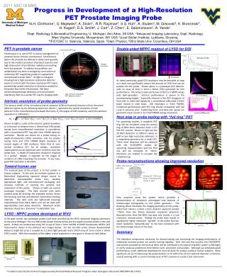
International Workshop on Radiation Imaging Detectors University of Glasgow, 25-29 July 2004. High spatial resolution measurement of depth-of-interaction of a PET LSO crystal. Outline. Background Basic concepts of PET

E N D
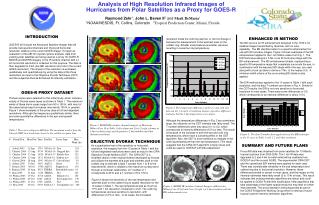
International Workshop on Radiation Imaging Detectors University of Glasgow, 25-29 July 2004 High spatial resolution measurement of depth-of-interaction of a PET LSO crystal
Outline Background Basic concepts of PET Challenges in small animal PET Our motivation Experimental Device under study Nuclear microprobe Microbeam irradiation of an LSO crystal Results Pulse height spectra and maps as a function of position from the detector Depth of interaction Conclusions
Basic concept of PET PET scanner of PET Center, University of Debrecen
Parallax error Schematic drawing of a PET device. The parallax error, influencing emission points far from the ring centre, is schematically shown.[2] A photon impinging on the entrance face of a detector with an oblique angle with respect to its axis can be detected not in that detector but in an adjacent one. Challenges in small animal PET Increased Spatial resolution is needed [1] Limitations of spatial resolution: positron range (~0.7mm tissue equivalent) non-collinearity of the annihilation photons parallax error [1] A. F. Chatziioannou: Molecular imaging of small animals with dedicated PET tomographs, European Journal of Nuclear Medicine 29 (1): 98-114 JAN 2002 [2] A. Braema, et al., Novel design of a parallax free Compton enhanced PET scanner, NIMA 525 (2004) 268.
The parallax error can be reduced or eliminated by measuring the interaction point of the photon along the detector. Depth of interaction DOI Different approaches to solve the DOI problem Improved and complex detector setup: (a) Phoswich detector approach (b) Stack approach (c) Detection of the light at the opposite bases of the scintillator Our aim was to investigate the effect of DOI on a LSO scintillator with high resolution
Experimental details • Sample • Commercially available LSO (Lutetium Oxyorthosilicate) • 1x1x10 mm3 crystal wrapped in a Teflon light reflector. • A ~0.70 mm wide, vertical cut along the Teflon enabled us • to irradiate the crystal itself. • Measurement • High resolution irradiation with a 2MeV He2+ beam at a • nuclear microprobe (beam size: 3x3 m2, ion rate: 300Hz) • Sequential scans of 1x1 mm2 areas along the 10 mm long crystal • DAQ with standard NIM
Nuclear microprobe components Accelerator Si(Li) Detector Computer Display X-Y Scan Coils Particle Detector Probe Formning Lens System B: analyzing magnet; C: condenser lens; S: beam steerer; Ob: object collimator; Ap: aperture collimator; P: vacuum pumps. M.B.H. Breese, D.N. Jamieson, P.J.C. King: Materials analysisusing a nuclear microprobe
LSO crystal 10mm Teflon wrapping 0.7mm 1mm 1mm LSO crystal Hamamatsu PhotomultiplierR5600 LSO Crystal Block Device under study
high counts low Results Counts/Channel Channel number Intensity map 1x1mm2 scan area, full energy window 128x128 pixel Pulse height spectrum of an 1x1mm2 area (512 channel)
0 0 5 5 1 1 6 6 counts 2 2 7 7 counts 3 3 8 8 4 4 9 9 5 5 10 10 Mean energy maps Intensity maps
500m Selected area spectra of 100mx1mm areas are extracted
Pulse height spectra of vertical areas (left centre and right ) between 5mm and 6 mm
Conclusions • Focused micro-ionbeamirradiation is a perspective technique • to study the position sensitive characterisics of a PET detector. • We have demonstrated that the interaction between the generated light and • detector can be studied with high lateral resolution (from m up to mm). • The mean value of the pulse height spectrum (~ position of the full energy peak) • is characteristic to the DOI. • Our results confirm previous gamma measurements on LSO crystal with similar • geometry and wrapping. • There is no difference between spectra collected from the left and right region • of the crystal. • Further advantage of the ion beam irradiation that there is no Compton-scattering • there is no Compton-background in the spectrum. • By varying the beam energy the penetration depth of the ions can be changed • giving opportunity for the real 3D light collection mapping. • (2mm projected range for 20 MeV protons)
Acknowledgements This work was supported by the Hungarian Scientific Research Fund Contract No. OTKA T34381 and National Office of Research and Technology Contract No. 1/0010/2002 .