Download
1 / 31
320 likes | 551 Views
Structure of the Heart and flow. Circuits of the heart. Systemic circuit- the left side of the heart pumps to the body (Contains oxygenated blood) Pulmonary circuit- the right side of the heart pumps to the lungs to oxygenate blood. The heart….

E N D
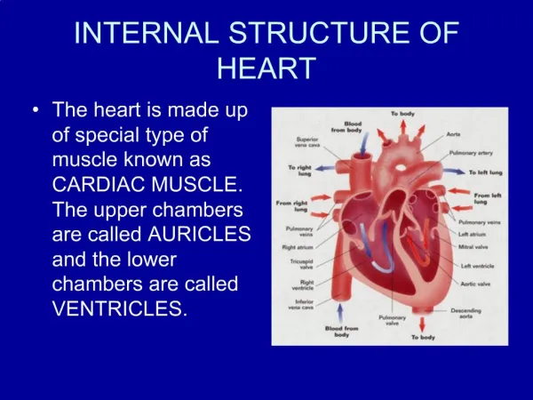
Circuits of the heart • Systemic circuit- the left side of the heart pumps to the body (Contains oxygenated blood) • Pulmonary circuit- the right side of the heart pumps to the lungs to oxygenate blood
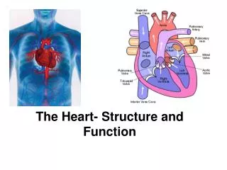
The heart…. • Location- in the middle of the chest directly below the sternum (breastbone) • Structure- the heart is enclosed in a sac, the pericardium, is a two layered wall. The space between the walls is filled with a lubricating fluid that allows the walls of the heart to slide over one another as the heart beats without friction • Comprised of cardiac muscle (myogenic muscle)- a type of muscle that is exclusive to the heart. It spontaneously contracts and relaxes quickly and without nervous system control • Supplied with blood by the coronary blood vessels • Requires over 10% of total O2 load of blood. This is greater than any other muscle of its size
Atria • Are known as the collection chambers • Receive blood passively and pump to the ventricles • Right Atria- receives blood from the body • Left Atria- receives blood from the lungs • The cardiac muscle that lines the walls of the atria are thin compared to the rest of the heart. • This is because the atria only need to pump the blood a short distance to the ventricles
Ventricles • Ventricles have a larger space and have thicker walls that are much more powerful than the atria. These thicker walls (especially the left ventricle) pump blood out to all the body organs. • Left is thicker because it pumps blood out to the whole body via the systemic circuit and the right is less thick because it pumps the shorter distance to the lungs
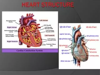
The two sides of the heart are separated by the septum. A thick muscular wall. • The pumping action of the heart is synchronized. The left and right atria pump together and the left and right ventricles pump together • Blood flows in one direction, with the help of valves 1) Atrioventricular valves (AV valves) • Separate atria and ventricles • Prevents blood from flowing from the ventricles back to the atria 2) Semilunar Valves (SL valves) • Separate ventricles from arteries • Prevent blood from flowing from the arteries back into the ventricles
Chordae Tendineae: White fibres that support the AV valves Mitral- Left AV Tricuspid- right AV Pulmonary -right SL Aortic – Left SL Heart murmur- unusual heart sound as a result of a leaky valve
SL valve https://www.youtube.com/watch?v=gSNty0jz0BU
Flow of blood • The circulatory system is comprised of a complex network of tubes that branch and rebranch to distribute blood and its contents to body cells • Largest ~ 3 cm • Smallest are 5-10µm • Blood flows from the; • Left side of Heart arteries arterioles capillaries venules veins back to the right side of the heart https://www.youtube.com/watch?v=PgI80Ue-AMo
Arteries • Carry blood away from the heart (toward body tissues) • Usually carries oxygenated blood (except the pulmonary artery) • High pressure • Thick walls and highly muscular • Oxygenated blood is pumped out of the left side of the heart through the aorta, a single large artery • It branches into the major arteries (see following diagram) • When the heart contracts it (squeezes in) it pushes blood into the arteries
The artery walls expand to accommodate for increased blood volume and pressure • The expansion you feel is your pulse • When the heart relaxes, the artery walls return to their original size and push the blood further along the blood vessels
Arterioles • Smaller than arteries • The size of arterioles can be controlled by the brain • VASODILATION- when the arterioles dilate to increase blood flow • When you are hot your arterioles dilate to move closer to the skin to lose heat. • VASOCONSTRICTION- when the arterioles contract to reduce blood flow to the skin to prevent loss of heat to the external environment. • When you are cold, the blood vessels constrict to move away from the surface of the skin
Capillaries • Arterioles branch into smaller blood vessels called capillaries when they reach the tissues. • A capillary network or capillary bed is an extensive network of blood vessels that supply oxygen and nutrients to every cell throughout the body tissues • Walls of the capillaries are 1 cell thick to allow for easy diffusion • Fluid and gas exchange –high to low concentration • As blood moves into the capillaries, pressure drops and blood slows for better diffusion
Capillaries cont… • Capillaries cannot be controlled by the nervous system • However, “pre-capillary sphincter muscles”can control blood flow into capillaries • If blood is needed in a particular area (i.e. muscles during exercise), the sphincter relaxes to allow blood flow to be increased • If blood is not needed in a particular area, the sphincters constrict to reduce blood flow • We would need 200L of blood if all our blood vessels were constantly open instead of the 4-5L
Venules and Veins • Return blood back to the heart, after passing through the capillary network and exchanging materials with the body • Capillaries merges with small vessels called venules • Venules merge into veins • Carry blood to the heart • Mostly deoxygenated and carry CO2 and other waste, except for the pulmonary vein • Vein walls are not as thick • Internal diameter is smaller and the pressure is significantly lower than in arteries
Venous return • How does blood under low pressure get back to the heart and move against gravity???? • Veins contain 1-way valves, that allow for blood to flow in one direction only • These veins are sandwiched between skeletal muscles. When these muscles contract, the veins are squeezed and the blood is forced in one direction back to the heart
Vericose Veins • Damaged valves can result in a condition known as varicose veins • Blood accumulates in the veins creating a bulge
http://www.sciencelearn.org.nz/Contexts/See-through-Body/Sci-Media/Animations-and-Interactives/Label-the-hearthttp://www.sciencelearn.org.nz/Contexts/See-through-Body/Sci-Media/Animations-and-Interactives/Label-the-heart • http://www.sporcle.com/games/g/heartanatomy - competition • http://www.purposegames.com/game/parts-of-the-human-heart-quiz -label competition