Download
1 / 17
220 likes | 527 Views
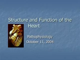
Heart Structure and Function. Lab III. Closed Circulatory System. Pulmonary Circulation: Carries O 2 -depleted blood away from the heart, to the lungs, and returns to the heart with O 2 rich blood.

E N D
Heart Structure and Function Lab III
Closed Circulatory System Pulmonary Circulation: Carries O2-depleted blood away from the heart, to the lungs, and returns to the heart with O2 rich blood Systemic Circulation: carries blood with O2 away from the heart to the rest of the body, and returns deoxygenated blood back to the heart
Circulation • Arteries • Heart pumps blood into them • Elastic and are able to expand and contract • Maintains proper blood pressure which allows flow to continue • Veins • Less elastic • Rely on skeletal muscle contractions to push blood • Has valves to prevent back-flow
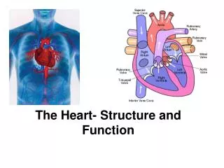
Structure Human Heart: • Separated into two pumps and four chambers • Right pump: right atrium and ventricle • Leads to lungs • Left pump: left atrium and ventricle • Leads to the body • Tip forms the apex Apex
Structure • Chambers connected by valves • Atrioventricular valve (AV): separates atria from ventricles • Tricuspid (R) • Bicuspid (L) • Semilunar: between ventricles and arteries • Aorta: carries blood away from the heart from the left ventricle • Pulmonary artery: carries blood from the right ventricle to the lungs • Open and close with pressure, prevent back-flow • Superior Vena Cava -> Right Atrium -> (Tricuspid) Valve -> Right Ventricle-> (Pulmonary Semilunar) Valve -> Pulmonary Artery -> Lungs -> Pulmonary Vein -> Left Atrium -> (Bicuspid) Valve -> Left Ventricle -> (Aortic Semilunar) Valve -> Aorta -> Body
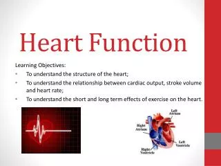
Today’s Lab: • Listen to heart sounds • Determine arterial and venous blood pressure • Analyze heart with EKG • Dissect sheep heart
Heart Sounds • Caused primarily by the shutting of valves which causes blood flow through the heart • Systole: Contraction of the heart • Atrial/Ventricular systole • Diastole: Period after systole when the heart fills with blood • Atrial/ventricular diastole
Heart Sounds • Two sounds associated with cardiac function • “Lub” • Closing of AV valves • Ventricles contract = systole • Long duration and low pitched • “Dub” • Closing of semilunar aortic valve (blood slamming against this) • Ventricles relax = diastole • Short duration and high pitched
Heart Sounds • Find your heart with a stethoscope • Listen for the “lub-dub” • Find where it is loudest – where is this on your chest?
Blood Pressure • Pressure exerted by circulating blood in arterial system against the walls of blood vessels • Ventricles contract and send a pressure wave through arteries = pulse • Sphygmomanometer or “sphygmos” • Measures the pressure wave in arterial blood pressure • Systolic pressure • Point of maximum pressure, during ventricular contraction • Sharp tapping sound • Diastolic pressure • Point of lowest pressure, during ventricular relaxation • Tapping becomes muffled
Blood Pressure • Arterial blood pressure • Systolic/Diastolic (mmHg) • Measured with a sphygmomanometer • Venous blood pressure • Much lower than arterial blood pressure • Measure mmH2O and convert to mmHg
EKG • Records electrical events in the Heart Electrical Activity of the Heart • Natural pacemaker in right atrium • Sinoatrial node (SA node) • Initiates the electrical sequence and causes the atria to contract • Electrical impulse from the SA node travels to the atrioventricular node (AV node) • Delays contraction of the ventricle so that the atria can empty their blood completely before the ventricles open Electrical current in the heart animation
EKG • 5 components • P, Q, R, S, T • P: Atrial contraction • QRS: Ventricular contraction • Simultaneous atrial relaxation • T: Ventricular repolarization • Recovery of ventricular muscle tissue to its resting state
Sheep Heart Dissection • View external anatomy • Try to identify any visible major blood vessels and arteries • Superior Vena Cava • Aorta • Pulmonary Artery • Pulmonary Veins • Internal anatomy • Identify chambers and valves
Be able to identify: -R/L atrium - Superior Vena Cava -R/L ventricle -Chordae tendinae -Aorta -Pulmonary Artery Explain the location of: -Pulmonary Vein -AV vales (Bicuspid/Tricuspid) Sheep Heart Dissection