Download
1 / 38
590 likes | 2.32k Views
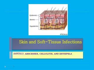
Soft Tissue Infections. Dr Cardinal. Objectives. Discuss the features of common skin infections as well as necrotizing skin infections including: Epidemiology Pathophysiology Clinical features Treatment. Necrotizing Soft Tissue Infections. Gas gangrene (Clostridial myonecrosis)

E N D
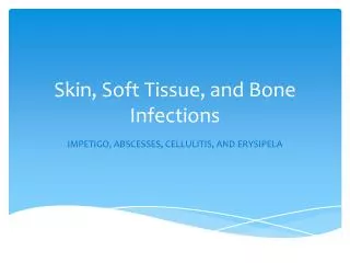
Soft Tissue Infections Dr Cardinal
Objectives • Discuss the features of common skin infections as well as necrotizing skin infections including: • Epidemiology • Pathophysiology • Clinical features • Treatment
Necrotizing Soft Tissue Infections • Gas gangrene (Clostridial myonecrosis) • Gas gangrene (Nonclostridial myonecrosis) • Streptococcal myositis • Necrotizing fasciitis (polymicrobial) • Necrotizing fasciitis ( Group A Streptococcus) • Necrotizing cellulitis
Epidemiology About 1000 cases reported to CDC yearly Pathophysiology Seven sp Clostridium C. perfringes – 80-95% Soil, GI tract, Female GU Release endotoxins Cardiodepressant Hydrolyzes cell membranes Gas Gangrene (Clostridial) C. perfringes
Clinical Incubation: 3 days Pain out of proportion Heaviness of affected part Edema, brown discoloration +/- crepitance Serosanguineous discharge Bullae Fever, tachycardia Confusion, sepsis Uterine myonecrosis after C-section Deepest of the necrotizing soft tissue infections Gas Gangrene (Clostridial)
Clinical X-Ray/CT: gas Treatment Resuscitation Antibiotics Pen G + Clindamycin Augmentin Imipenem Unasyn Surgical debridement Hyperbaric oxygen therapy (HBO) Gas Gangrene (Clostridial)
Pathophysiology Mixed infections Aerobic / anaerobic Bacteria by prevalence: Enterococcus Staphylococcus Alpha-Streptococci E. coli Klebsiella Proteus Bacteroides Gas Gangrene (Nonclostridial) E. Faecalis in blood culture
Clinical Similar to Clostridial except: Pain at onset less Delayed presentation (2-10 days) Mortality 43% Gas Gangrene (Nonclostridial)
Treatment Broad spectrum antibiotics Aerobic gram + / - Anerobes Unasyn Timentin Zosyn Imipenem Floroquinolone added if fresh water infection suspected Surgical debridement HBO Gas Gangrene (Nonclostridial)
Rare muscle infection Group A Streptococcus Usually S. pyogenes “flesh eating bacteria” Epidemiology Age 20-50 Otherwise healthy Streptococcal Myositis
Clinical Difficult to distinguish from other myonecrosis No gas production High rate of bacteremia Toxic shock syndrome ~ 4-6 hours after admission Mortality: 80-100% Streptococcal Myositis
Treatment Aggressive management of shock Early vasopressors in shock Antibiotics IV Pen G / Clindamycin IVIG 2g/kg Neutralizes exotoxins Surgical debridement Streptococcal Myositis
Necrosis of subQ and fascia Does not spread to muscle as clostridial / nonclostridial myonecrosis Also grouped into tabloid term “flesh eating bacteria” Necrotizing Fasciitis (Polymicrobial)
Epidemology 10-20 cases per 100,000 people DM, PVD, IVDA, smoking Pathophysiology Mixed aerobic / anerobic Average 4.4 organisms per infection Usually antecedent trauma / bite Bacteremia 25-30% Mortality 25-50% Necrotizing Fasciitis (Polymicrobial)
Clinical Pain out of proportion Erythematous / edematous Discoloration, vesicles Fever, tachycardia May progress rapidly Crepitus as late finding “Finger test” Necrotizing Fasciitis (Polymicrobial)
Treatment Aggressive resuscitation Avoidance of vasopressors Antibiotics as in myonecrosis Surgery HBO Necrotizing Fasciitis (Polymicrobial)
Epidemiology 10-20 cases per 100,000 Mortality 20-60% Increased risk with: Varicella lesions NSAID use Necrotizing Fasciitis (Group A Streptococcus)
Clinical Same as polymicrobial except: No gas formation More rapid progression More prone to bacteremia Toxic shock more common Necrotizing Fasciitis (Group A Streptococcus)
Treatment Initial broad spectrum Abx Narrowed to PCN and Clindamycin after culture Clindamycin Synergistic to PCN Suppresses toxin formation Promotes phagocytosis Suppresses PCN binding protein HBO of little use (aerobic organism) Necrotizing Fasciitis (Group A Streptococcus)
Most superficial necrotizing soft tissue infection Involves skin and subQ Pathophysiology Associated with antecedent trauma / bite Common in skin “popping” Usually polymicrobial Clostridium most common C. perfringens in trauma C. septicum in malignancy and spontaneous infection Necrotizing Cellulitis
Clinical Pain at site. Less than seen in deeper infections Eccymotic or necrotic center Vesicles or blebs possible Crepitence +/- Mild or no systemic symptoms Necrotizing Cellulitis
Treatment Surgical debridement (usually curative) Broad spectrum abx Necrotizing Cellulitis
Local soft tissue inflammation from bacterial invasion Epidemiology 1.3% of ER vistis 61% Male, Mean age 46 Usually extremities Only 5% have predisposing factors Cellulitis
Pathophysiology Adults – Staph/Strep spp. most common Children – H. influenza most common Diabetics – consider Enterobacteriaceae Most bacterial organism cleared from body with 12 hours. Majority of symptoms are from host immune response Cellulitis
Clinical Local inflammation, tenderness, warmth, erythema, induration Lymphangitis uncommon – concerning Bacteremia uncommon in healthy hosts Cellulitis
Treatment outpatient Dicloxacillin Macrolide Azithromycin Clarithromycin Augmentin Treatment inpatient Involving head or neck IV abx Cellulitis
Superficial cellulitis with lymphatic involvement Usually from Group A Streptococcus Antecedent trauma / bite or dermatoses Lower extremities now most common Erysipelas
Clinical Abrupt onset High fever, chills, malaise and nausea prodrome Area of erythema with burning develops over next 2 days Red, shiny, hot plaque, sharply demarcated Erysipelas
Treatment usually inpatient Pen G IV Nafcillin Rocephin Augmentin Imipenem in severe Erysipelas
Pathophysiology Requires loss of skin integrity Usually caused by common colonizers Scalp/trunk/extremities Staph aureus, epidermidis Intriginous/perineal E. coli, P. mirabilis Axilla P. mirabilis Cutaneous Abscesses
Clinical Swelling, tenderness, erythema Fluctuance, induration, drainage Systemic spread unusual in healthy Lymphadenitis, fever Cutaneous Abscesses
Specific abscesses Bartholin gland abscess Paronychia and felons Hidradenitis suppurativa Infected sebaceous cyst Perirectal abscess Pilonidal abscess Cutaneous Abscesses
Staphylococcal abscesses Continuum of severity Folliculitis Furuncle (boil) Carbuncle Cutaneous Abscesses
Treatment Incision and drainage Wide excision if necessary (furuncle) Antibiotics Always in high risk groups Controversial in healthy persons Cutaneous Abscesses
Mycotic infection Sporothrix schenckii Common in soil and on vegetation Thermally dimorphic Traumatic inoculation Sporotrichosis
Clinical Incubation 3 weeks Crusted ulcer or plaque at site Local lymphadenitis common Skip lesions along lymphatics Rarely systemic Meningitis Pulmonary Sporotrichosis
Diagnosis Clinically / History Fungal cultures Tissue biopsy Treatment Itraconazole for 6 months Sporotrichosis