Download
1 / 97
1.01k likes | 1.06k Views
Learn about bacterial and viral skin infections, differentiate staphylococci from streptococci, list common pyogenic skin infections, and explore immune system responses. Discover treatment options and management strategies.

E N D
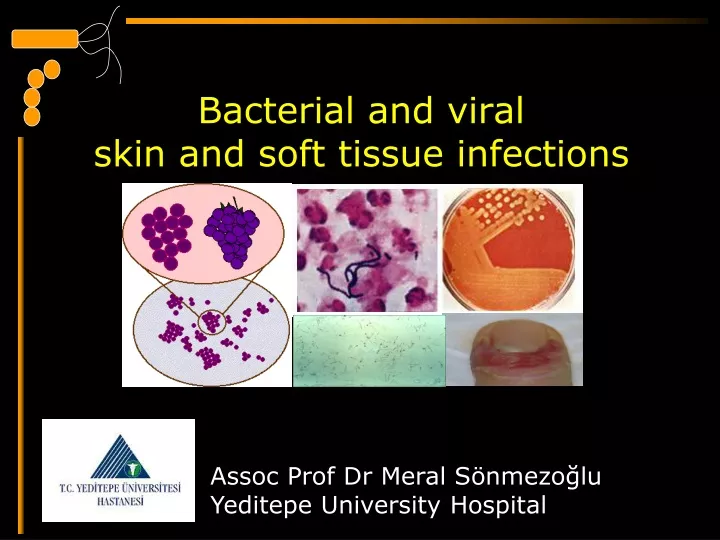
Bacterial and viral skin and soft tissue infections Assoc Prof Dr Meral Sönmezoğlu Yeditepe University Hospital
Learning objectives Differentiate staphylococci from streptococci, and name several skin infections caused by each. List the causative agent, mode of transmission, and clinical symptoms of folliculitis, furunculitis, ,impetigo, erisipelas and viral skin infections
Common pyogenic skin infections Folliculitis Furunculosis Carbuncles Paronychia Impetigo Cellulitis Erysipelas Surgical wound infection Other soft tissue infections Tetanus Gas gangrene Necrotising facsiitis Staphylococcal Scalded Skin Syndrome Dermatophyte infections Osteomyelitis Septic arthritis Overview
Skin infections • Soft tissue infections are common • Generally mild-modest severity • Sign and symptoms of toxicity: • Fever/hypothermia • Tachycardia (>100 beats/min) • Hypotension • CBC, Cr, CPK, CRP, blood culture
Skin infections • Potentially severe deep soft tissue; • Pain • Violaceous bullae • Cutaneous hemorrhage • Skin sloughing • Skin anesthesia • Rapid progression • Gas in the tissue • Imaging
SKIN INFECTIONS • Impetigo, erysipelas, cellulitis • Necrotising infections • Infections following animal and human bites • Infections associated with animal contact • Surgical site infections (SSI) • Infections in the immunocompromised host
Staphylococcus aureus Gram-positive cocci in clusters Catalase-positive Coagulase-positive Streptococcus pyogenes Gram-positive cocci in chains Catalase-negative Group A beta-haemolytic streptococcus Staphs & Streps
Immune System • Innate Immunity • Epithelial cells • Dendritic cells • Macrophages • Natural killer cells • Neutrophils
The Structure of Human Skin • Perspiration and sebum contain nutrients • Salt inhibits microbes • Lysozyme hydrolyzes peptidoglycan • Fatty acids inhibit some pathogens Figure 21.1
Microbial Diseases of the Skin • Exanthem: Skin rash arising from another focus of the infection • Enanthem: Mucous membrane rash arising from another focus of the infection
Skin Lesions Figure 21.2
Folliculitis • Infection of hair follicles • usually pustular folliculitis • Clinical presentation • follicle-centred pustules • e.g. in scalp, groin, beard & moustache (sycosis barbae) • Mostly (95%) due to Staphylococcus aureus • Treatment: oral flucloxacillin
Pyogenic skin infections • Furunculosis • form of deep folliculitis. • Carbuncle • multiple abscesses in close apposition with interconnecting sinuses. • Acute paronychia • Skin infection arising from nail • Treatment • Oral flucloxacillin
Furuncules and carbuncles • Furuncules are infections of hair follicle • Caused by S. aureus • Suppuration extends through the dermis into the subcutaneous tissue (small abcess) • Inflammatory nodule and overlying pustule • Several adjacent follicles produce carbuncle (neck, diabetes)
folliculitis staphylococcal pustulosis furuncle carbuncle
Management • Small furuncles: drainage • Larger furuncles and all carbuncles: incision and drainage • Systemic antibiotic unnecesary (fever) • Recurrent furuncules: eradication of carriage (mupirocin) • Clindamycin (150 mg 1x1 5 days)
Impetigo • Discrete purulent lesions • Prevalent during summer months • Peak incidence among 2-5 years • Colonization the unbroken skin precede the development, (personal hygiene, minor trauma, bites) • 10 days
Impetigo • Superficial infection • usually staphylococcal (nose) • but can also involve Streptococcus pyogenes (urt) • Friable, golden crusts over erythematous skin. • Treatment • Topical fucidin or mupirocin 7-10d • Oral flucloxacillin or erythomycin • if widespread or unresponsive
Nonbullous Lesions of Impetigo Figure 21.4
Cellulitis • Diffuse, acute, spreading skin infection • Involves deeper dermis and subcutanous fat • Edema, redness, heat, regional LAP
Cellulitis • Diffuse parenchymal inflammation without necrosis or localisation of pus • Often seen as erythematous halo around a wound • Commonly caused by S. aureus • Less common causes: S. pyogenes, C. perfringens • Treatment • oral pen V + flucloaxcillin • or co-amoxiclav • If severe may require i-v treatment
Erysipelas • Lesions raised above the level of skin • Affects upper dermis (superficial lymphatics) • Well-demarcated cellulitis with fever and malaise • acute streptococcal infection • bacteremia common • upper dermal oedema lifts epidermis except where staked down by hair follicles or sweat glands • leads to the typical “peau d'orange” appearance • Treatment: penicillin V
Staphylococcal toxic shock syndrome Desquamation of the skin, especially on the palms and soles, occurs 10 to 21 days after the onset of the disease. Another late finding is a pruritic, generalized maculopapular eruption developing on days 9 to 13. Patients can lose their nails and hair after 4 to 16 weeks, with resolution of these abnormalities by 5 to 6 months.
Surgical Wound Infection • Features: • induration • fever • erythema • leakage of pus • may have infection in absence of pus (e.g. streptococcal cellulitis, gas gangrene, infected burns) • Treatment: debridement and antibiotics (flucloxacillin ± benzylpenicillin)
Gas Gangrene • infectious disease emergency • Caused by exotoxin-producing Clostridium perfringens • usually after direct inoculation of contaminated, ischaemic wound
Gas GangreneClinical features • Myonecrosis, gas production, and sepsis • rapid onset and progression to toxaemia and shock • crepitus, brawny oedema • foul-smelling discharge, brown skin discoloration, bullae, dead muscle • infection can advance 1 ,5 cm per hour! • pain out of proportion to physical findings • Mortality greater than 25%
Gas gangrene • Diagnosis: • Clinical • Radiological • gas within the fascial planes • Microbiological • Gram-positive rods in tissues, culture of C. perfringens • Treatment • fasciotomy, debridement, amputation • antibiotics (penicillin and metronidazole) • hyperbaric oxygen (?)
Necrotising Fasciitis • Similar condition to gas gangrene, but usually lacking gas production • Caused by S. pyogenes and/or S. aureus, often in combination with anaerobes • mortality rate of 30-50% • Treatment • debridement, amputation • antibiotics
Staphylococcal Scalded Skin Syndrome • seen in infants, young children, immuno-compromised • epidermolytic toxin released into the blood stream from localised S. aureus infection • causes widespread superficial exfoliation
VIRAL SKIN INFECTIONS Meral Sonmezoglu, MD. Assoc Professor of Infectious Dıseases
VIRAL SKIN INFECTIONS • Several common childhood viral infections; • Rubeola (Measles) • Rubella (German measles) • Varicella (Chickenpox) • Erythema infectiosum (fifth disease) • Erythema subitum (roseola) • Echovirus-adenovirus
OTHER VIRAL SYNDROMES • Pityriasis rosea (HHV 6,7?) • Hand, foot and mouth disease • Gianotti Crosti syndrome • Haemorragic fevers • Smallpox • HIV infections • Rickettsial diseases
LOCAL SKIN VIRAL INFECTIONS • Herpes simplex • Herpes zoster • Vesicular stomatitis • Molloscum contagiosum • Viral warts
CLIN. MAN. OF VIRAL INFECTIONS • Erythema multiforme • Erythema nodosum • Kawasaki disease
MEASLES • Measles, also known as rubeola, is a disease caused by a virus, a member of the family paramyxoviridae, genus Morbillivirus • Measles is spread through respiration (contact with fluids from an infected person's nose and mouth, either directly or through aerosol transmission), and is highly contagious—90% of people without immunity sharing a house with an infected person will catch it.