Download
1 / 2
20 likes | 51 Views
An open letter from Dr. Jane Edmond for eye care providers regarding management of BBSOAS with regards to vision. Download Dr. Edmondu2019s letter and share with all eye care providers.

E N D
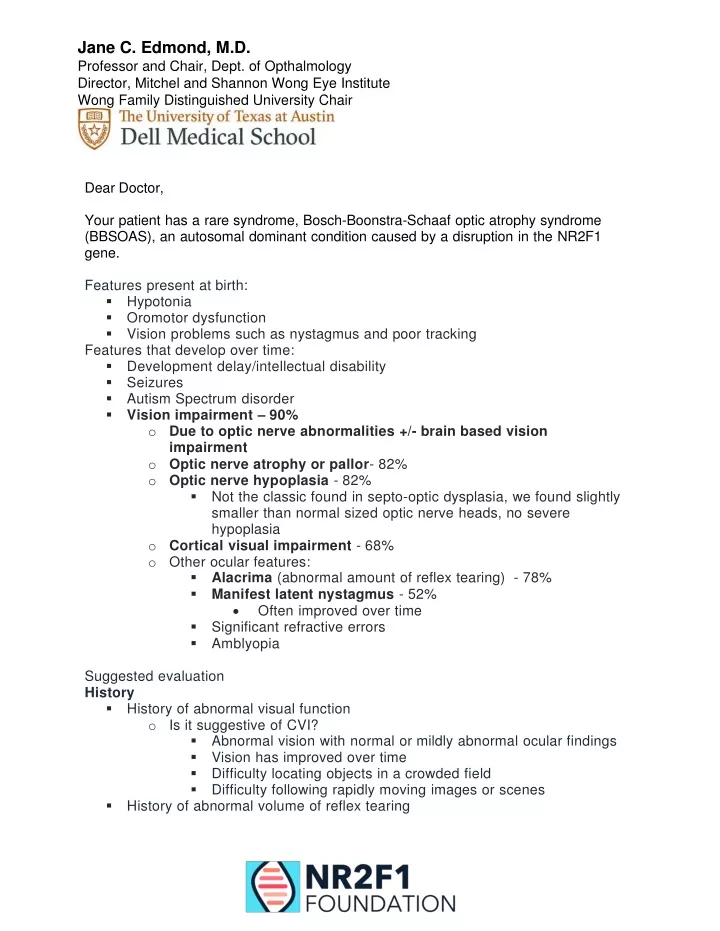
Jane C. Edmond, M.D. Professor and Chair, Dept. of Opthalmology Director, Mitchel and Shannon Wong Eye Institute Wong Family Distinguished University Chair Dear Doctor, Your patient has a rare syndrome, Bosch-Boonstra-Schaaf optic atrophy syndrome (BBSOAS), an autosomal dominant condition caused by a disruption in the NR2F1 gene. Features present at birth: ▪ Hypotonia ▪ Oromotor dysfunction ▪ Vision problems such as nystagmus and poor tracking Features that develop over time: ▪ Development delay/intellectual disability ▪ Seizures ▪ Autism Spectrum disorder ▪Vision impairment – 90% oDue to optic nerve abnormalities +/- brain based vision impairment oOptic nerve atrophy or pallor- 82% oOptic nerve hypoplasia - 82% ▪ Not the classic found in septo-optic dysplasia, we found slightly smaller than normal sized optic nerve heads, no severe hypoplasia oCortical visual impairment - 68% o Other ocular features: ▪Alacrima (abnormal amount of reflex tearing) - 78% ▪Manifest latent nystagmus - 52% • Often improved over time ▪ Significant refractive errors ▪ Amblyopia Suggested evaluation History ▪ History of abnormal visual function o Is it suggestive of CVI? ▪ Abnormal vision with normal or mildly abnormal ocular findings ▪ Vision has improved over time ▪ Difficulty locating objects in a crowded field ▪ Difficulty following rapidly moving images or scenes ▪ History of abnormal volume of reflex tearing
Exam ▪ Visual acuity (test which is appropriate for patient’s age and understanding) o Teller acuity cards, Allen pictures, LEA, HOTV, Snellen o Color vision (indicator of optic nerve function) ▪ External exam o Presence of a lacrimal gland? o Manifest latent nystagmus? ▪ Pupil reactivity o Poorly reactive, APD? ▪ Visual fields o Nonspecific abnormalities due to optic nerve abnormalities or CVI ▪ Intraocular Pressure o Glaucoma was not a finding in our pts ▪ Anterior segment o Dry eye findings? ▪ Optic nerve o Mild hypoplasia? ▪ Normal optic nerve diameter • ~2.5 disc diameters fit in between the border of the optic nerve and the fovea ▪ Mild hypoplasia • >2.5 disc diameters fit in between the border of the optic nerve and the fovea o Optic atrophy?, mild or severe Suggested In-office testing, if able: ▪Schirmer’s testing ▪ Visual fields o Confrontation, if able, automated perimetry ▪ Fundus photography to document optic nerve health o Document distance between optic nerve border and fovea (rule out mild hypoplasia ▪ OCT o RNFL to document health of the optic nerve Thank you for helping our BBSOAS families, Jane C. Edmond, M.D. For more information, visit NR2F1 Foundation: https://nr2f1.org/