Download
1 / 20
500 likes | 1.88k Views
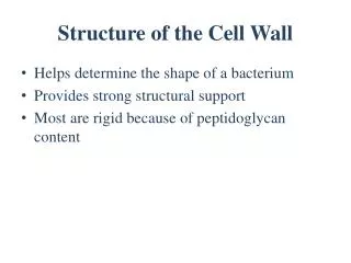
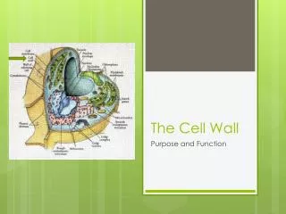
Cell wall structure. I. Function. A Prevent cell rupture due to osmotic pressure B Provides Shape C Anchors flagella. II. Clinically important. Site of antibiotic action B. Virulence factor that causes disease. III Components. A. Peptidoglycan B. Peptides and proteins

E N D
I. Function A Prevent cell rupture due to osmotic pressure B Provides Shape C Anchors flagella
II. Clinically important • Site of antibiotic action B. Virulence factor that causes disease
III Components A. Peptidoglycan B. Peptides and proteins C. Phospolipids D. Polysaccharides
IV What is a Peptidoglycan Layer? A rigid lattice structure made of long polymer of repeating disaccharide sugars that are bond together by polypeptide chains • Polymer of disaccharide sugar • Made of alternating N-acetylglucosamine (NAG) and N-acetylmurmic acid (NAM) • Form long rods that are refered to as the "carbohydrate backbone”
The Peptidoglycan Layer (cont.) • The carbohydrate backbones are held in place laterally by peptide cross bridges • Peptide cross bridge binds NAG • The carbohydrate backbones are held together vertically by tetrapeptides • Tetrapeptides bind to NAM
Gram Positive Cell Walls • Cell wall is made up of many peptidoglycan layers (very thick and rigid) • Contain teichoic acids • Lipoteichoic acids spans peptidoglycan layer and links to the plasma membrane. • Wall teichoic acid links to peptidoglycan layer. • Functions of teichoic acid • Movement of cations • Suport wall durring cell growth • Strong antigenic determinant
Gram Negative Cell Walls • Outer membrane is unique in all of biology. Creates a periplasmic space. • Peptidoglycan layer inside the periplasmic space • Protein receptors that regulate chemotaxis.
Gram Negative Cell Walls (cont.) 2. Outer membrane composed of • Phospholipid bilayer: Protects the cell against antibiotics and other chemicals which attack the peptidoglycan layer • Lipopolysaccharide layer: • O polysccharide (Strongist antigenic determinant). • Lipid A (endotoxin). Extremely toxic in minute amount • Porin proteins: Channel by which nutrients and waste pass through the cell wall. • Lipoproteins: Proteins that connect the outer membrane and the plasma membrane
Gram Stain • Primary stain (crystal violet) • Wash • Mordant (Iodine) • Wash • Decolorizer (alchol/ acid wash) • Counter stain (Safranin) • Gram pos = primary stain sticks (crystal violet) • Gram neg. = Primary stain fails to bind. These cells are visualized by adding the counter stain (safranin)
Movement of Materials Across Membranes • Passive Processes • Simple diffusion: movement of molecules from areas of high concentration to area of low concentration. • Facilitated diffusion: Molecules combines with a plasma membrane protein called a transporter. • Osmosis: The movement of water across a selectively permeable membrane. High concentration to low concentration
Movement of Materials Across Membranes • Active Transport: The cell uses energy (ATP) to move substance across the plasma membrane. • Moves substance from low concentration to high concentration • Depends on transporter protein • Group translocation: Substance is altered once it is across the membrane to make it impermeable to the membrane.