Download
1 / 30
310 likes | 367 Views
This guide provides methods for isolating and identifying Staphylococci and Streptococcus, essential Gram-positive cocci in clinical microbiology. Learn about Gram-staining, culture techniques, direct identification tests, and key tests like Mannitol Fermentation, Coagulase Test, and DNase Test. Discover distinct characteristics of Staphylococcus aureus, Staphylococcus saprophyticus, and β-haemolytic Streptococcus groups. Explore identification steps based on Lancefield groups, including Streptococcus pyogenes (Group A), Streptococcus agalactiae (Group B), and more. Get insights into group-specific antigens and key diagnostic tests like hemolysis patterns, antibiotic sensitivity tests, and CAMP test. Understand the unique features of Streptococcus pneumoniae (Pneumococcus) and Enterococci for accurate laboratory identification.

E N D
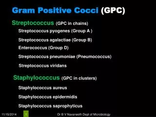
Identified method for Staphylococci • Gram-stain • Isolation and culture • Pure culture • Directidentification
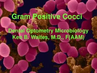
Staphylococci are Gram-positive cocci, typically arranged in clumps or Grape-like clusters
Directidentification • The mannitol fermentation test. • The Coagulase Test • The DNase Test • Novobiocin (NB) disc
The mannitol fermentation test. • Inoculate the bacteria into a mannitol micro-tube,incubate at 370C for 18h.S.aureus will ferment mannitol to produce acid,which causes the medium to turn yellow.
The Coagulase Test • Coagulase is an enzyme converting fibrinogen into fibrin promoting blood clotting. • It might be a virulence factor with the coagulated blood around the bacteria protecting them from the immune system. • Coagulase-negative strains are often as pathogenic as coagulase-positive strains.
The DNase Test • Inoculate DNase agar plates with a loop so that the growth is in plaques about 1 cm in diameter.I • ncubate at 370C overnight. • Flood the plate with 1 N hydrochloric acid.Clearing around the colonies indicates DNase activity. • The hydrochloric acid reacts with unchanged deoxyribonucleic acid to give a cloudy precipitate. • A few other bacteria,e.g. Serratia,may give a positive reaction.
Blood agar with a novobiocin (NB) disc Staphylococcus aureus Growing on Blood Agar Staphylococcus saprophyticus Growing on Blood Agar
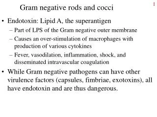
β haemolytic steps: Lancefield groups • Historically • A, B, C, F and G group specific antigens cell wall polysaccharide • group D and Enterococci lipoteichoic acid moiety Not always β haemolytic • Now: 18 different groups of strep: from A-H, and from K-T
(β) Haemolytic Streptococcus groups • S. pyogenes • Group A • S. agalactiae • Group B (occasionally α or none) • Streptococcus equismilis • Group C (occasionally α or none) • Streptococcus bovis • Group D (α or none, occasionally β) • Streptococcus anginosus • Group F & G
Gp A strep: S. pyogenes Laboratory ID • Gram +ve • Catalase • Haemolysis • small, round, grey colonies • β haemolysis • zone is large • Gp A antigen
Isolation and Identification of group A beta hemolytic (Streptococcus pyogenes) • on Blood agar. Streptococcus pyogenes produces a zone of beta hemolysis around 2-3mm in diameter surrounding each colony • Sensitivity to the antibiotic bacitracin • Group A Strep is senstive, other b haemolytic streps are resistant.
Group B: S. agalactiae • S. agalactiae • Identification • Catalase negative • Appearance on BA • Larger colonies than Gp A • Small zone of hemolysis • Latex agglutination Gp B • Bacitracin resistant • CAMP positive • Growth on Mac (weak)
Presumptive ID Tests - CAMP • The “CAMP” test (acronym for developers) is used to differentiate S. agalactiae (GBS), which is positive, from other beta hemolytic strep, all of which are negative • “CAMP factor” is a soluble hemolysin produced by GBS that combines in a synergistic way with a similar hemolysin of S. aureus to form an arrowhead zone of clearing. See image • The staph is streaked perpendicularly to the unknown strep. After appropriate incubation time an “arrowhead-shaped” clear zone of hemolysis will appear.
S. agalactiae • Synergistic haemolysis • observed between betahemolytic • Staphylococcus • aureus and group B • streptococci. • Positive reaction: arrowhead haemolysis pattern
α – Haemolytic streps: Viridans streps • Not groupable by Lancefield groups • No group specific CHO • Streptococcus pneumoniae • Primary human pathogen • S.sanguis • S.mitis • S.mutans • S.salivarius
The Pneumococcus (Streptococcus pneumoniae) 1: Isolation on Blood agar • Pneumococci frequently require enriched media and increased CO2 tension for initial isolation. • They are usually isolated on Blood agar and incubated in a candle jar (a closed container in which a lit candle is placed to remove O2 and increase CO2 ) at 37C. On Blood agar, colonies appear small, shiny, and translucent. • They are surrounded by a zone of alpha hemolysis 2: Optochin sensitivity • Pneumococci are the only streptococci that are sensitive to the drug optochin(ethylhydrocupreine hydrochloride). 3: Gram stain:gram-positive, diplococci
The Genus Enterococcus • Enterococci are gram-positive streptococci, typically occurring in pairs and short chains, that are normal flora of the intestinal tract. Enterococci responsible for a variety of opportunistic infections in humans, and serologically belong to Lancefield group D streptococci. • On Bile Esculin agar • Unlike most bacteria, the enterococci will grow in the presence of the bile salts in the medium. • They hydrolyze the esculin, producing esculetin which reacts with the iron salts in the medium turning the agar black
Laboratory ID: Enterococcus • white colonies • alpha and no haemolysis (rarely beta) • Also grow on MacConkey • Resembles S. pneumoniae in Gram stains • Bile esculin positive • Grows on Mac