Download
1 / 55
630 likes | 961 Views
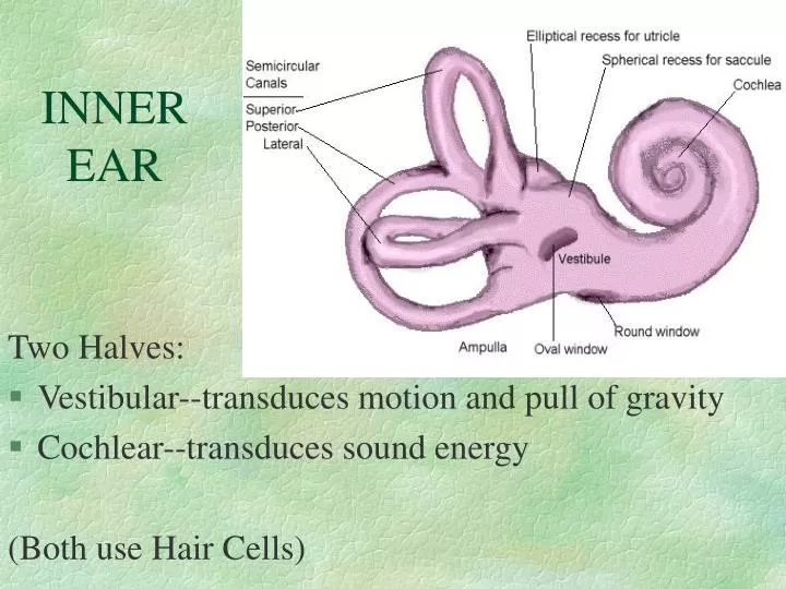
INNER EAR. Two Halves: Vestibular--transduces motion and pull of gravity Cochlear--transduces sound energy (Both use Hair Cells). Subdivision into spaces containing endolymph (blue) , and spaces containing perilymph (red). The Endolymphatic Sac. Termination of vestibular aquaduct

E N D
INNER EAR Two Halves: • Vestibular--transduces motion and pull of gravity • Cochlear--transduces sound energy (Both use Hair Cells)
Subdivision into spaces containing endolymph (blue), and spaces containing perilymph (red)
The Endolymphatic Sac • Termination of vestibular aquaduct • Outside of temporal bone; next to dura mater lining of the brain • Thought to maintain endolymphatic volume/pressure
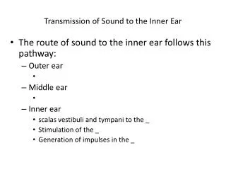
Cochlea is Divided into 3 “Scala” • Scala Vestibuli • Reissner’s Membrane • Scala Media • Basilar Membrane • Scala Tympani • Helicotrema - the opening between 2 outer Scala
Fluids filling the Inner Ear • Perilymph- in S. Vestibuli and S. Tympani • High Sodium / Low Potassium concentrations • Low Voltage (0 to +5 mV) • Endolymph- in S. Media • High Potassium / Low Sodium concentrations • High Positive Voltage (80 mV)
Cross-Section of the Cochlea Third Turn Second Turn First Turn
I = Inner Hair Cells P = Pillar Cells O = Outer Hair Cells D = Deiter’s Cells
IHCs, OHCs And Their Stereocilia • OHCs (at top) • 3, 4 or 5 rows • Approx 12,000 cells • 10 to 90 microns • V- or W-shaped ranks of stereocilia • 50 to 150 stereocilia per cell • IHC (at bottom) • 1 or 2 rows • Approx 3,500 cells • 35 microns • straight line ranks of stereocilia • 50 to 70 stereocilia per cell
Cochlear Functions • Transduction- Converting acoustical-mechanical energy into electro-chemical energy. • Frequency Analysis-Breaking sound up into its component frequencies
Transduction- • Inner Hair Cells are the true sensory transducers, converting motion of stereocilia into neurotransmitter release. Mechanical Electro-chemical • Outer Hair Cells have both forward and reverse transduction-- Mechanical Electro-chemical Mechanical Electro-chemical
Frequency Analysis - the Traveling Wave • Bekesy studied cochleae from cadavers, developed the Traveling Wave theory • 1. Response always begins at the base • 2. Amplitude grows as it travels apically • 3. Reaches a peak at a point determined by frequency of the sound • 4. Vibration then dies out rapidly
Bekesy’s Theory describes Passive Mechanics • Based on work in “dead” cochleae • Highly damped -- not sharply tuned • Active Undamping occurs in live and healthy cochleae • Like pumping on a swing--adds amplitude
The Active Component • Improves Sensitivity for soft sounds • Improves frequency resolution
Frequency Tuning Curves Show these Effects = plots of response threshold as a function of frequency They have a characteristic shape • sharp tip (shows best sensitivity at one freq) • steep high frequency tail • shallow low frequency tail
Tuning Curves Passive Only Active + Passive
More on Tuning & Tuning Curves: • Seen for basilar membrane, hair cells, nerve cells • Frequency of “tip” is called the CHARACTERISTIC FREQUENCY
OHC Length and CF High Freqs Low Freqs
Hair Cell Activation • Involves Ion Flow into cell • Through channels in the stereocilia • Bending stereocilia causes # of open channels to change. • Toward Modiolus = Fewer channels open • Away from Modiolus = More open
Ion Channels are opened by “TIP LINKS” • Tip Links connect tip of shorter stereocilia to the side of a stereocilium in the next taller row • Bending toward taller rows pulls tip links • Bending toward shorter rows relaxes tip links
Resting (or Membrane) Potentials • Inner Hair Cell = - 45 mV • Outer Hair Cell = - 70 mV
Stereocilia bent toward tallest row • Potassium flows into cell • Calcium flows into cell • Voltage shifts to a less negative value • More neurotransmitter is released
Synapse Basics • Pre-Synaptic cell contains vesicles • Gap between cells is Synaptic Cleft • Post synaptic cell may show darkened area adjacent to membrane
4 Types of Cochlear Neurons • INNER HAIR CELLS • Multiple (10 to 20) Afferent synapses • (Efferents synapse on afferent dendrites) • OUTER HAIR CELLS: • Large Efferent synapses engulf base of cell • Small (& not very active) Afferent synapses
Inner hair cells • Synapse at the base with up to 20 afferent neurons • “Divergence” • Efferents synapse on afferent dendrites under IHCs
Afferent neurons have their cell bodies in the Spiral Ganglion (4)
Cochlear Potentials: • Resting Potentials: voltages which exist without external stimulation e.g., Endolymphatic Potential, Cell Membrane Potential • Stimulus-Related Potentials: voltages occurring in response to sounds We’ll talk about 3 of these from the cochlea
Cochlear Microphonic • Least valuable from a clinical standpoint. • Is an alternating current (AC) response that mirrors the waveform of low to moderately intense sound stimuli • Appears to arise from outer hair cells in the basal-most turn of the cochlea