Download
1 / 16
270 likes | 1.26k Views
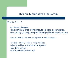
Chronic lymphocytic leukemia (1). Is characterised by the accumulation of nonproliferating mature-appearing lymphocytes in the blood, marrow, lymph nodes, and spleen In most cases, the cells are monoclonal B lymphocytes that are CD5+ T cell CLL can occur rarely.

E N D
Chronic lymphocytic leukemia (1) • Is characterised by the accumulation of nonproliferating mature-appearing lymphocytes in the blood, marrow, lymph nodes, and spleen • In most cases, the cells are monoclonal B lymphocytes that are CD5+ • T cell CLL can occur rarely
Chronic lymphocytic leukemia (2) • Is the most common form of leukemia in North America and Europe, but is extremely rare in the Orient • Typically occurs in older patients, with the highest incidence being in those aged 50 to 55 years • Affects men twice as often as women
Etiology (1) • The cause of CLL is unknown • There is increased incidence in farmers, rubber manufacturing workers, asbestos workers, and tire repair workers • Genetic factors have been postulated to play a role in high incidence of CLL in some families
Etiology (2) • Cytogenetics • clonal chromosomal abnormalities are detected in approximately 50% of CLL patients • the most common clonal abnormalities are: • trisomy 12 • structural abnormalities of chromosomes 13, 14 and 11 • patients with abnormal karyotypes have a worse prognosis • Oncogenes • in most cases of CLL is overexpressed the proto-oncogene c-fgr 9a member of the src gene family of tyrosine kinases
Clinical findings (1) • Approximately 40% of CLL patients are asymptomatic at diagnosis • In symptomatic cases the most common complaint is fatigue • Less often the initial complaint are enlarged nodes or the development of an infection (bacterial)
Clinical findings (2) • Most symptomatic patients have enlarged lymph nodes (more commonly cervical and supraclavicular) and splenomegaly • The lymph nodes are usually discrete, freely movable, and nontender • Hepatomegaly may occure • Less common manifestation are infiltration of tonsils, mesenteric or retroperitoneal lymphadenopathy, and skin infiltration • Patients rarely present with features of anemia, and bruising or bleeding
Laboratory findings (1) • The blood lymphocyte count above 5,0 G/L • In most patients the leukemic cells have the morphologic appearance of normal small lymphocytes • In the blood smears are commonly seen ruptured lymphocytes (“basket” or “smudge” cells) • Careful examination of the blood smear can usually differentiate CLL, and the diagnosis can be confirmed by immunophenotyping
Laboratory findings (2) • Clonal expansion of B (99%) or T(1%) lymphocyte • In B-cell CLL clonality is confirmed by • the expression of either or light chains on the cell surface membrane • the presence of unique idiotypic specificities on the immunoglobulins produced by CLL cells • by immunoglobulin gene rearrangements • typical B-cell CLL are unique in being CD19+ and CD5+ • Hypogammaglobulinemia or agammaglobulinemia are often observed • 10 - 25% of patients with CLL develop autoimmune hemolytic anemia, with a positive direct Coombs’ test • The marrow aspirates shows greater than 30% of the nucleated cells as being lymphoid
The diagnostic criteria for CLL 1) A peripheral blood lymphocyte count of greater than 5 G/L, with less than 55% of the cells being atypical 2) The cell should have the presence of Bcell-specific differentiation antigens (CD19, CD20, and CD24) and be CD5(+) 3) A bone marrow aspirates showing greater than 30% lymphocytes
Differential diagnosis • Infectious causes • bacterial (tuberculosis) • viral (mononucleosis) • Malignant causes • B-cell • T-cell • leukemic phase of non-Hodgkin lymphomas • Hairy-cell leukemia • Waldenstrom macroglobulinemia • large granular lymphocytic leukemia
Investigations • Pretreatment studies of patients with CLL should include examination of: • complete blood count • peripheral blood smear • reticulocyte count • Coomb’s test • renal and liver function tests • serum protein electrophoresis • immunoglobulin levels • plasma 2 microglobulin level • If available immunophenotyping should be carried out to confirm the diagnosis • Bone marrow biopsy and cytogenetic analysis is not routinely performed in CLL
Staging (1) • Rai Classification for CLL • 0 - lymphocytosis (>5 G/L) • I - lymphocytosis + lymphadenopathy • II - lymphocytosis + splenomegaly +/-lymphadenopathy • III - lymphocytosis + anemia (Hb <11g%) +/-lymphadenopathy or splenomegaly • IV - lymphocytosis + thrombocytophenia (Plt <100G/L) +/- anemia +/-lymphadenopathy +/- splenomegaly
Staging (2) • Binet Classification for CLL • A. < 3 involved areas, Hb > 10g%, Plt > 100G/L • B. > 3 involved areas, Hb > 10g%, Plt > 100G/L • C. - any number of involved areas, Hb < 10g%, Plt < 100G/L
Rai classification stage median survival (years) 0 >10 I > 8 II 6 III 2 IV < 2 Binet classification stage median survival (years) A > 10 B 7 C 2 Prognosis
Markers of poor prognosis in CLL • Advanced Rai or Binet stage • Peripheral lymphocyte doubling time <12 months • Diffuse marrow histology • Increased number of prolymphocytes or cleaved cells • Poor response to chemotherapy • High 2- microglobulin level • Abnormal karyotyping
Treatment • Treatment is reserved for patients with low- or intermediate risk disease who are symptomatic or have progressive disease (increasing organomegaly or lymphocyte doubling time of less than 12 months) and patients with high -risk disease • Alkylating agents (chlorambucil, cyclophosphamide) • Nucleoside analogs (cladribine, fludarabine) • Biological response modifiers • Monoclonal antibodies • Bone marrow transplantation • And systemic complications requiring therapy • antibiotics • immunoglobulin • steroids • blood products