Download
1 / 36
380 likes | 1.05k Views
Chapter 12. Circulatory Disturbances. Learning Objectives. Venous thrombosis: causes and effects Pulmonary embolism: pathogenesis, clinical manifestations, diagnostic techniques Arterial thrombosis: causes and effects

E N D
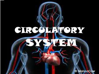
Chapter 12 Circulatory Disturbances
Learning Objectives • Venous thrombosis: causes and effects • Pulmonary embolism: pathogenesis, clinical manifestations, diagnostic techniques • Arterial thrombosis: causes and effects • Edema and factors regulating fluid circulation between capillaries and interstitial tissue (hydrostatic pressure, capillary permeability, osmotic pressure, open lymphatic channel) • Hypercoagulable state • Shock: pathogenesis and treatment
Intravascular Blood Clots • Normally, blood does not clot within the vascular system • Pathogenesis of intravascular clotting • 1. Slowing or stasis of blood flow • 2. Blood vessel wall damage • 3. Increased coagulability of blood • Thrombus: an intravascular clot; can occur in any vessel or within the heart • Embolus: a detached clot carried into pulmonary or systemic circulation; plugs vessel of smaller caliber than diameter of clot, blocking blood flow and causing necrosis • Infarct: tissue necrosis from interruption in blood flow
Embolism (1 of 5) • From blood clots, fat, air, amnionic fluid, and foreign particles • Fat embolism • Following severe bone fracture that disrupts fatty bone marrow and surrounding adipose tissue • Emulsified fat globules sucked into veins and carried into lungs, obstructing pulmonary capillaries • If it reaches systemic circulation, eventually blocks small vessels in brain and other organs
Embolism (2 of 5) • Air embolism • Large amount of air sucked into circulation from lung injury due to a chest wound • May be accidentally injected into circulation • Air carried into right heart chambers and prevents filling of heart by returning venous blood • Heart unable to pump blood and individual dies rapidly of circulatory failure
Embolism (3 of 5) • Amnionic fluid embolism • Devastating complication of pregnancy • Amnionic fluid enters maternal circulation through a tear in fetal membranes • Fetal cells, hair, fat, and amniotic debris fluid block maternal pulmonary capillaries causing severe respiratory distress • Thromboplastic material in fluid activates coagulation mechanism leading to disseminated intravascular coagulation syndrome
Embolism (4 of 5) • Foreign particulate matter embolism • Various types of particulate material • May be injected by substance users that crush and dissolve tablets intended for oral use • Material injected intravenously and is trapped within small pulmonary blood vessels • Symptoms of severe respiratory distress
Embolism (5 of 5) • Septic emboli • Thrombi form in pelvic vein following uterine infection • Bacteria invade thrombi • Emboli from infected thrombus travel to lungs, causing pulmonary infarct • Bacteria in clot invade pulmonary infarct causing lung abscess
Venous Thrombosis • Predisposing factors to clot formation in leg veins • Prolonged bed rest • Cramped position for an extended period • Impaired “milking action” of leg musculature that normally promotes venous return resulting in stasis of blood in veins • Varicose veins or any condition preventing normal emptying of veins • Outcome • Leg swelling from partial blockage of venous return in leg • Pulmonary embolism
Pulmonary Embolism (1 of 5) • Clinical manifestations depend on size of embolus and where it lodges in the pulmonary artery • Large pulmonary emboli may completely block main pulmonary artery or major branches obstructing blood flow to lungs • Lung not infarcted due to collateral blood flow from bronchial arteries (from descending aorta) that interconnect with pulmonary arteries via collateral channels • Cyanosis and shortness of breath due to inadequate oxygenation of blood
Pulmonary Embolism (2 of 5) • Large pulmonary emboli • 1. Right side of heart becomes distended • 2. Pulmonary artery becomes overdistended with blood, causing increased pulmonary pressure • 3. Left ventricle unable to pump adequate blood to brain and vital organs • 4. Systemic blood pressure falls and patient may go into shock
Pulmonary Embolism (3 of 5) • Small pulmonary emboli • Small emboli may pass through main pulmonary arteries, becoming impacted in peripheral arteries supplying lower lobes of the lungs • Raises pulmonary pressure and inadequate collateral circulation • Affected lung segment undergoes necrosis; wedge-shaped pulmonary infarct • If infarct develops: dyspnea, pleuritic chest pain, cough, and expectoration of bloody sputum due to leakage of blood from infarcted lung tissue into bronchi
Pulmonary Embolism (4 of 5) • Diagnosis • Chest X-ray: detects infarct but not the embolus • Radioisotope lung scans: detects abnormal pulmonary blood flow caused by embolus • Pulmonary angiogram (gold standard): detects blocked pulmonary artery • Computed tomography (CT) scan: detects pulmonary embolus indicated by obstructed flow of contrast medium, information comparable to pulmonary angiography without requiring insertion of catheter in pulmonary artery
Chest X-ray Lung Scan
Pulmonary Embolism (5 of 5) • Treatment • Anticoagulants: heparin initially followed by coumadin • Thrombolytic drugs if with massive embolus • Angioplasty (balloon or stent to widen vein) • Thrombectomy (clot extraction surgery) • General supportive care
Arterial Thrombosis (1 of 2) • Stasis is not a factor due to rapid blood flow and high intravascular pressure • Main cause: injury to vessel wall from arteriosclerosis, causing ulceration, roughening of arterial with thrombi formation • Blocks blood flow • Coronary artery: myocardial infarction • Major leg artery: gangrene • Cerebral artery: stroke
Arterial Thrombosis (2 of 2) • Intracardiac thrombosis • Clot forms • Within atrial appendages: heart failure • Surfaces of heart valves: valve injury • Wall of left ventricle: myocardial infarction • May dislodge into systemic circulation and cause infarction: spleen, kidneys, brain
Thrombosis by Increased Coagulability (1 of 2) • 1. Rise in coagulation factors following surgery or injury • 2. Estrogen in contraceptive pills stimulates synthesis of clotting factors • 3. Hereditary gene mutations • Mutation of gene that codes for factor V results in abnormal factor V Leiden: more resistant to inactivation, prolonged activity, increased coagulability • Mutation of gene regulating prothrombin synthesis • Risk for venous thrombosis increases as prothrombin level rises
Thrombosis by Increased Coagulability (2 of 2) • 4. Thrombosis in patients with cancer, from increased platelets and coagulation factors • Predisposes to both arterial and venous thromboses • Hypercoagulability due to rapid release of thromboplastic materials into circulation from tumor deposits • Platelets and coagulation factors consumed faster than can be replenished, leading to bleeding • Large tumors release thromboplastic material slowly but continuously; production of coagulation factors exceeds destruction leading to hypercoagulability
Edema • Accumulation of fluid in interstitial tissues, first noted in ankles and legs • Results from disturbance of extracellular fluid circulation between capillaries and interstitial tissues • Pitting edema: pit or indentation formed when edematous tissue is compressed with the fingertips • Hydrothorax: fluid accumulates in pleural cavity • Ascites: fluid accumulates in peritoneal cavity
HP =hydrostatic pressure • Due to fluid pressing against a wall • “Pushes” • In capillary (HPc) • Pushes fluid out of capillary • 35 mm Hg at arterial end and 17 mmHg at venous end of capillaryin this example •In interstitial fluid (HPif) • Pushes fluid into capillary • 0 mm Hg in this example Arteriole Venule Interstitial fluid Capillary Net HP—Net OP (17—0)—(26—1) Net HP—Net OP (35—0)—(26—1) OP =osmotic pressure • Due to presence of nondiffusible solutes (e.g., plasma proteins) • “Sucks” •In capillary (OPc) • Pulls fluid into capillary • 26 mm Hg in this example •In interstitial fluid (OPif) • Pulls fluid out of capillary • 1 mm Hg in this example Net HP 35 mm Net OP 25 mm Net OP 25 mm Net HP 17 mm NFP (net filtration pressure) is 10 mm Hg; fluid moves out NFP is ~8 mm Hg; fluid moves in Figure 19.17
Net Filtration Pressure (NFP) • NFP—comprises all the forces acting on a capillary bed • NFP = (HPc—HPif)—(OPc—OPif) • At the arterial end of a bed, hydrostatic forces dominate • At the venous end, osmotic forces dominate • Excess fluid is returned to the blood via the lymphatic system
Pathogenesis of Edema • Increased capillary permeability • Causes swelling of tissues with acute inflammation • Increase in capillary permeability from some systemic diseases • Low plasma proteins • Excess protein loss (kidney disease) • Inadequate synthesis (malnutrition) • Increased hydrostatic pressure • Heart failure • Localized venous obstruction • Lymphatic obstruction
Factors Regulating Fluid Flow Between Capillaries and Interstitial Tissue • 1. Capillary hydrostatic pressure: force pushing fluid from capillaries into extracellular space • 2. Capillary permeability: determines ease of fluid flow through capillary endothelium • 3. Osmotic pressure: water-attracting property of a solution; exerted by proteins in the blood (colloid osmotic pressure) that attract fluid from interstitial space back into the capillaries • 4. Open lymphatic channels: collect fluid forced out of the capillaries by the hydrostatic pressure and return fluid into circulation
Edema © Courtesy of Leonard Crowley, M.D./University of Minnesota Medical School
Edema © Courtesy of Leonard Crowley, M.D./University of Minnesota Medical School
Circulatory Shock • Any condition in which • Blood vessels are inadequately filled • Blood cannot circulate normally • Results in inadequate blood flow to meet tissue needs
Circulatory Shock • Hypovolemic shock: results from large-scale blood loss • Vascular shock: results from extreme vasodilation and decreased peripheral resistance • Cardiogenic shock results when an inefficient heart cannot sustain adequate circulation
Shock (1 of 2) • Low blood flow/pressure to adequately supply body with blood; potentially life-threatening; circulating blood volume < capacity of vascular system • Categories according to pathogenesis • Hypovolemic shock: low blood volume • Cardiogenic shock: reduced cardiac output • Septic shock: excessive vasodilatation secondary to release of microbial toxins and inflammatory mediators • Anaphylactic shock: excessive vasodilatation from release of inflammatory mediators
Shock (2 of 2) • Prognosis depends on early recognition and rapid appropriate treatment • Drugs that promote vasoconstriction • Use of intravenous fluids or blood to restore blood volume secondary to fluid loss or hemorrhage • Treat underlying cause