Download
1 / 23
240 likes | 408 Views
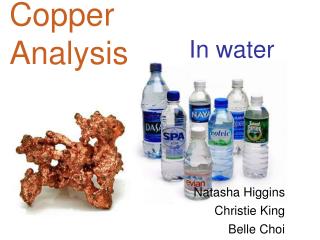
Analysis of Copper in PDB files. Joana Pinto July 2009. Objective. Analyse the structures of Copper containing proteins and look for errors Keep making protein structures more and more accurate Relying on improved software and better knowledge of protein structure. PDB Files Errors.

E N D
AnalysisofCopper inPDB files Joana Pinto July 2009
Objective • Analysethestructures of Copper containing proteins and look for errors • Keep making protein structures more and more accurate • Relyingon improved software and better knowledge of protein structure
PDB Files Errors Difficulty in distinguish certain atoms Errors are kept because it’s not a curated database.
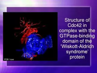
Ions in Proteins • Iron, Zinc, Calcium, Magnesium, Chlorine or Iodine, etc. • Copper • Two stable oxidation states • [Ar] s1d10 • Copper proteins classification
Type I Copper Proteins S - Methionine Cysteine - S Cu N - Histidine N - Histidine Blue Proteins 2 Histidines, 1 Cysteine and 1 Methionine There are about 50.000 proteins in this family
Type II Copper Proteins N - Histidine Cu N - Histidine X N - Histidine 3 Histidines + Histidine or H2O or Methionine or Cysteine Colorless
Type II Copper Proteins - PHM • Peptidylglycine α-Hydroxylating Monooxygenase
Type II Copper Proteins - PHM CuA CuB
Type III Copper Proteins N - Histidine N - Histidine O N - Histidine Cu Cu N - Histidine O N - Histidine N - Histidine Binuclear centers: 3 Histidines and O2
Type III Copper Proteins- Hemocyanin *Oxygenated state 3,6 Å *Deoxygenated state 4,6 Å * Not validated by us
Cu+ versus Cu2+ Reduced form - cuprous Oxidized form - cupric • [Ar] 4s03d10 • Soft ion Soft ligands (S/P) • C.N: 2, 3 and 4 • [Ar] 4s03d9 • Hard ion Hard ligands (N/O) • C.N: 4, 5 and 6
Materials and Methods • PDB Files: • X-ray Crystallography • Resolution better than 2.0 Å
Materials and Methods • WHAT IF: • Calculations • SKPNOO • GRSION • Distances plot • YASARA: • Pictures
Results– Regular tetrahedrals for Cu+ and Cu2+ 13 Cu+ ions (spread over 8 PDB files) and 149 Cu2+ ions (spread over 94 PDB files) distances plot
Results–Distancesplot Average positions for all the intervenient residues in the regular tetrahedrals difference between the average positions is of 0.6 Å, being this value close to the error expected in the X-ray structure determination
Results–Flipping Histidines * Guss, J.M., Harrowell, P.R., Murata, M., Norris, V.A., Freeman, H.C. (1986). Crystal Structure Analysis Of Reduced (Cu I) Poplar Plastocyanin At Six pH Values. J.Mol. Biol 192 (1986): 361-387 • When superposed, 5PCY* and 6PCY* show no differences in its structures besides: • a small change in a Proline residue, and a rotation of an Imidazole ring by 180˚ about Cβ - Cγ.
Results–Flipping Histidines 5PCY 6PCY No reason for the flipping of the Histidine At a resolution of both 5PCY (1.80 Å) and 6PCY (1.90 Å) the Pro puckering tends to be unobservable
Conclusions *Belle, C., Rammal, W., Pierre, J.L., (2005). SulfurLigation in Copper Enzymes and Models. Journal of Inorganic Biochemistry 99: 1929-1936 **Taylor, M.K., Stevenson, D.E., Berlouis, L.E.A., Kennedy, A.R., Reglinski J. (2006). Modeling the Impact of Geometric Parameters On The Redox Potential Of Blue Copper Proteins. Journal of Inorganic Biochemistry 100: 250-259 • Too few data • Coordination Numbers: • For Cu+ and Cu2+: most often seen C.N. is 4, 5 and 3. • Contradicting some older studies* • No differences between the structures of Cu+ and Cu2+ • Contradicting some older studies**
Conclusions • For coppers with C.N. 4, the most common geometry is tetrahedral • Copper proteins classifications should be revised, for example, Type II classification is ambiguous. • Some older publications should be reviewed
FutureWork • What distinguishes between both oxidation states for copper? • Are the differences too small to be detected? • Are there any differences at all? • One of the possibilities to be studied would be the protonation states for Histidine and Cysteine or other factors like protein bond
Thank You Questions?