Download
1 / 44
440 likes | 668 Views
Somatic Nervous System & Special Senses. Chapter 12 . Special Senses. Part B. Sciences of Special Senses. Ophthalmology – is the science that deals with the eye and its disorders Otorhinolaryngology – deals with all the other senses. . Olfaction: Smell. Sense of smell

E N D
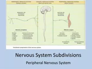
Somatic Nervous System & Special Senses Chapter 12
Special Senses Part B
Sciences of Special Senses • Ophthalmology – is the science that deals with the eye and its disorders • Otorhinolaryngology – deals with all the other senses.
Olfaction: Smell • Sense of smell • Requires 10 million to 100 million receptors
Structures • Olfactory epithelium • Located in the upper portion of the nasal cavity • Consists of three types of cells • Olfactory receptors • Supporting cells • Basal stem cells
Olfactory Receptors • Are the stimulated by olfactory hairs, which project from knob-shaped tip of the olfactory receptor • Odorants (chemicals in the air) stimulate the olfactory hairs • Short life span – only about 1 month • See nose hairs are good
Supporting Cells • Columnar epithelial cells of the mucous membrane lining of the nose • Provide physical support, nutrients, and electrical insulation for the olfactory receptors
Basal Cells • Cells that are responsible for producing new olfactory receptors
Olfactory glands • Responsible for the production of mucus that moistens the surface the surface of the olfactory epithelium and serves as a solvent for inhaled odorants.
Stimulation of Olfactory Receptors • Unique because adaptation to a chemical is very rapid • You may smell something bad but over time if you remain in that environment you won’t smell it anymore.
Hyposmia • Reduced ability to smell, affects over half of those over the age of 65 and 75% over 80. • Caused by head injury, Alzheimer’s disease or Parkinson’s disease • Also by certain drugs, such as antihistamines, or steriods
Gustation: Taste • Five primary tastes • Sour • Sweet • Bitter • Salty • Umani
Umami • Describe as a “savory” taste
Taste Buds • Taste buds – are the location of receptors for taste • Taste buds are found on the papillae, which are the bumps on the tough • Number of tastes bud decrease with age • Location • Mostly on the tongue • Some on the roof of mouth, throat and epiglottis
Tastants • Chemicals that activate the gustatory receptors
Stimulation • Tastants are dissolved in saliva which then allows them to enter the pores and come into contact with the gustatory hairs. • Stimulation of hairs causes an electrical impulse to be sent to the brain
Vision • More than half the sensory receptors in the human body are found in the eye • The largest part of the cerebral cortex is devoted to vision
Careers Associated with vision • Ophthalmologist – physician who specializes in the diagnosis and treatment of eye disorders with drugs, surgery and corrective lenses • Optometrist – has a doctorate of optometry and is licensed to test the eyes and treat visual defects b y prescribing corrective lenses • Optician – technician who fits, adjusts and dispenses corrective lenses using the prescription supplied by an ophthalmologist or optometrist
Accessory structures of the eye • Accessory structures – are eyebrows, eyelashes, eyelids, muscles the move eyeballs and lacrimal (tear) apparatus
Eyelashes and Eyebrows • Protect the eyeballs from foreign objects, perspiration, and direct rays from sun
Eyelids • Upper and lower • Shade the eye during rest • Protect eyes from excessive light • Protect eyes from foreign objects • Spread lubricating secretions over the eyeballs by blinking
Eye Muscles • Six muscles control the movement of the eyeball • Right, left, up, down and diagonally
Lacrimal apparatus • Group of glands, ducts, sacs that produce and drain lacrimal fluid as tears • Lacrimal glands (one for each eye) • About the size and shape of an almond and are responsible for secreting lacrimal through the lacrimal ducts to the eyeballs • Tears can also pass through the nasolacrimal duct, which allows lacrimal to drain into your nasal cavity
Lacrimal • Is a watery solution containing salts, some mucus, and a bacteria killing enzyme called lysozyme
Layers of the eyeball • Adult eyeball measures about 2.5 cm across • Three layers • Fibrous Tunic • Vascular Tunic • Retina
Fibrous Tunic • Outer coat of the eyeball • Consists of • Cornea – transparent fibrous coating that colors the iris (colored portion of your eye) • Also helps focus light • Sclera – is the “white” of the eye, dense connective cove • Gives the shape of the eye, makes it more rigid and protects inside of eyeball • Conjunctiva – epithelial layer, covers the anterior surface of eyeball and lines inner surface of eyelid
Corneal Transplant • If your cornea is damaged it can be replaced with a donor cornea
Vascular Tunic • Middle Layer of the eye • Composed of: • Choroid – thin membrane that lines most of the internal surfaces of the sclera • Contains many blood vessels that nourish the retina • Ciliary body – consists of • ciliary processes, which are folds on the inner surface of the ciliary body whose capillaries secrete a fluid called aqueous humor and the • ciliary muscles, a smooth muscle that alters the shape of the lens for viewing objects up close or at a distance • Lens – transparent structure that focuses light rays onto the retina • Constructed of many layers of elastic protein fibers • Attached to ciliary muscles by zonular fibers
Iris and pupil • Part of the vascular tunic • Iris • Colored portion of the eye • Muscle that regulates the amount of light let into the eye • Pupil • Hole in the center of the eye through which light enters
Retina • Portion that lines posterior of the eye about ¾ of the eyeball • Consists of two layers • Neural layer • Pigmented layer
Neural Layer • Consists of three distinct layers • Photoreceptor layer • Bipolar cell layer • Ganglion cell layer
Photoreceptor layer • Two types of cells • Rods – allow us to see shades of gray in dim light (6 million) • Cones – allow us to see color (120 million) • Found in central fovea in the center of the macula lutea
Pigmented Layer • A sheet of melanin is located between the choroid and the neural part of the retina • Melanin absorbs stray light, helping to keep the image sharp and clear
Pathway • Photoreceptor layer to • Bipolar cell layer to • Ganglion cell layer to • Optic nerve
Interior of Eyeball • Two cavities divided by the lens • Anterior cavity • Vitreous cavity
Anterior cavity • Contains aqueous humor that helps maintain the shape of the eye and supplies oxygen and nutrients to the lens and cornea.
Vitreous Chamber • Contains the vitreous body, which helps maintain the shape of the eyeball and keeps the retina attached to the choroid • Vitreous body – develops in embryonic life and is not replaced • Choroid – provides blood supply and absorbs scattered light
Intraocular pressure • Pressure in the eye • Maintains the shape of the eye • Keeps the retina pressed against choroid • Normal pressure is 16mm of Hg