Download
1 / 26
270 likes | 561 Views
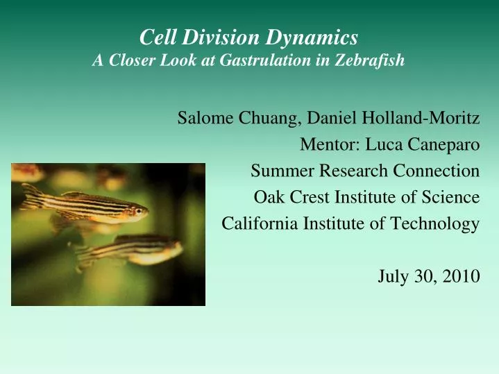
Cell Division Dynamics A Closer Look at Gastrulation in Zebrafish. Salome Chuang, Daniel Holland-Moritz Mentor: Luca Caneparo Summer Research Connection Oak Crest Institute of Science California Institute of Technology July 30, 2010. Cell Division: A Crucial Biological Process. Normal:

E N D
Cell Division Dynamics A Closer Look at Gastrulation in Zebrafish • Salome Chuang, Daniel Holland-Moritz • Mentor: Luca Caneparo • Summer Research Connection • Oak Crest Institute of Science • California Institute of Technology • July 30, 2010
Cell Division: A Crucial Biological Process • Normal: • Growth • Development • Tissue repair • Abnormal: • Cancer http://tainano.com/chin/Molecular%20Biology%20Glossary.htm
Spindle Poles Our Project: Dynamics of Cell Division in Embryos End of Cell Division Beginning of Cell Division
Difficulties of Studying Cell Division in Live Embryos • Ethical concerns (humans) • Opaque embryos • Development Rate • Production Rate • Manipulation • Storage/Care • External vs. Internal • Development Images from Kidscientist.com and odec.ca
Zebrafish: An Ideal Model for Studying Embryonic Cell Division • Very common for use in research because of: • Rapid reproductive rate • Transparent embryos • Rapid development • Easily cared for and contained • Easy to induce and screen for mutations • Genome almost fully sequenced Bar: 250 µm Images from focusonnature.be
A Brief History on Zebrafish George Streisinger Established zebrafish as an ideal subject for imaging and manipulation. Very small species of minnow Native to the Ganges region of India Named for distinctive stripes Since then zebrafish have been popularized as a model for studying vertebrate development: Cell biology and development Organ system development Genetic studies Image from www.zfin.org
Stages of Zebrafish Development Key: =Injection stage =Imaging stage
Cell Division in Dorsal Region of Embryos Dorsal Posterior
Significance of the Dorsal Region Dorsal region becomes... Nervous system Application: Cell orientation could have an impact on brain and nervous system development at an early age Cell imaging provides better insight into.... Embryonic development Cell division mechanisms and signaling in vertebrates
Shield Stage to Gastrulation: Trends • Tissue differentiation • Dorsal region: Definite pattern • Cells divide along the anterior-posterior plane Concha, Adams, 1998
Spindle Poles Visualizing Cell Division Dynamics End of Cell Division Beginning of Cell Division
Labeling of Cell Structures to Study Cell Division Dynamic • Red: Nucleus • Green: Cytoskeleton
MethodsGenetic Engineering of Fluorescent Embryos • mRNA codes for fluorescent protein in cytoskeleton and nucleus DNA RNA Protein • Synthetic mRNA produced for injection • mRNA injected into embryos at one cell stage Image from protein-dynamics.l001.de
MethodsImaging of Embryos Using Confocal Microscopy • Confocal microscope detects fluorescence • Embryos oriented to image dorsal region • Anterior and posterior marked • Imaged over a period of 4 hours
Point assigned to each half of separating chromosomes Program tracks and measures the angle of division relative to the anterior-posterior plane and dorsal-ventral plane Analysis • Special computer program tracks division alignment Image Courtesy of L. Caneparo
Results We observed an active rotation of the cells to align prior to division. Before After
Future Directions • Further Analysis • Quantify angle/character of rotation • Statistically analyze data • Investigate cell signaling patterns • Compare cell division in other vertebrate models • Explore implications on neural development
Acknowledgements • Special Thanks to: • Luca Caneparo • Fraser Lab • Sherry Tsai • James Maloney • California Institute of Technology • Howard Hughes Medical Institute • Siemens Foundation • Our families