Download
1 / 43
470 likes | 1.3k Views
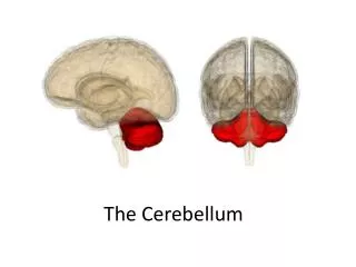
The Cerebellum. Position. Lies above and behind the medullar and pons and occupies posterior cranial fossa. Cerebellum. External features. Consists of two cerebellar hemisphere united in the midline by the vermis. External features. Three peduncles

E N D
Position • Lies above and behind the medullar and pons and occupies posterior cranial fossa Cerebellum
External features Consists of twocerebellar hemisphere united in the midline by the vermis
External features Three peduncles • Inferior cerebellar peduncle -connect with medulla and with spinal cord, contain both afferent and efferent fibers • Middle cerebellar peduncle -connect with pons, contain afferent fibers • Superior cerebellar peduncle -connect with midbrain, contain mostly efferent fibers
External features • Tonsil of cerebellumtwo elevated masses on inferior surface of hemispheral portion just nearby foramen magnum
Lobs • Two deep fissures • Primary fissure • Posterolateral fissure • Three lobs • Flocculonodular lobe 叶flocculus and nodule • Anterior lobe • Posterior lobe Corpus of cerebellar
Lobs Anterior lobe corpus of cerebellar Primary fissure Posterior lobe Flocculonodular lobe Posterolateral fissure
Internal structures Gray matter • Cerebellar cortex • Cerebellar nuclei • Dentate nucleus • Fastigial nucleus • Interposed nucleus • Emboliform nucleus • Globose nucleus White matter-medullary center
Internal structures Fastigial nucleus Cerebellar cortex Globose nucleus Dentate nucleus Emboliform nucleus medullary center
Three functional divisions • Vestibulocerebellum • Archicerebellum • Flocculonodular lobe • Spinocerebellum • Paleocerebellum • Vermis and intermediate zone • Cerebrocerebellum • Neocerebellum • Lateral zone Intermediate zone Vermis Lateral zone Flocculonodular lobe
Connections and function of cerebellum Vestibulocerebellum • Connections • Afferents: receive input from vestibular nuclei and primary vestibular • Efferents: projects to the vestibular nucleus → vestibulospinal tract and medial longitudinal fasciculus → motor neurons of anterior horn • Function: involved in eye movements and maintain balance
Connections and function of cerebellum Spinocerebellum • Connnection • Afferents: receive somatic sensory information via spinocerebellar tracts
Efferents: • Vermis projects to the fastigial nucleus → vestibular nuclei and reticular formation → vestibulospinal tract and reticulospinal tract → motor neurons of anterior horn • Intermediate zone projects to the interposed nuclei • Contralateral red nucleus → rubrospinal tract →motor neurons of anterior horn • Contralateral VI →cerebral cortex→ coticospinal tract→motor neurons of anterior horn • Function: play an important role in control of muscle tone and coordination of muscle movement on the same side of the body
Connections and function of cerebellum Cerebrocerebellum • Connection • Afferents: receives input from the cerebral cortex via a relay in pontine nuclei • Efferents: projects to dentate nucleus → VI → primary motor cortex → corticospinal tract → motor neurons of anterior horn • Function: participates in planning movements
小脑的分叶和功能 • 前庭小脑-古小脑 - 绒球小结叶 维持身体姿势平衡和协调眼球运动 • 脊髓小脑-旧小脑 包括蚓垂、蚓锥体和前叶 控制运动中的肢体远端肌的肌张力和协调 • 大脑小脑-新小脑:协调肢体的随意运动,使运动更精确
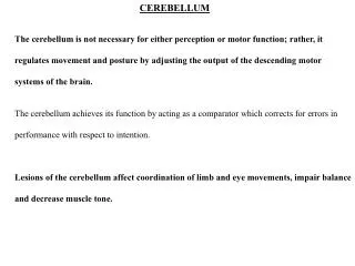
小脑损伤的临床表现 • 小脑是运动的重要调节中枢,有大量的传入和传出联系。大脑皮质发向肌肉的运动信息和执行运动时来自肌肉和关节等的信息,都可传入小脑。小脑经常对这两种传来的神经冲动进行整合,并通过传出纤维调整和纠正各有关肌肉的运动,使随意运动保持协调。此外,小脑在维持身体平衡上也起着重要作用。它接受来自前庭器官的信息,通过传出联系,改变躯体不同部分肌肉的张力,使肌体在重力作用下,作加速或旋转运动时保持姿势平衡。此外,据研究,小脑对内脏机能活动也有一定作用。小脑损伤引起的功能障碍是同侧性的。小脑受损伤后功能障碍主要表现为:肌张力低下,肌肉弛缓,如出现小腿呈钟摆样反射;随意运动发生障碍,表现为运动的速度、范围、力量和方向不准确,如步态失调,动作笨拙;平衡障碍,如躯体不易维持直立姿势,而向受损侧倾斜;植物性神经系统功能障碍,如尿失禁。
Position • Position: Lies between midbrian and cerebrum, almost entirely surrounded by cerebral hemisphere
Subdivision • Doral thalamus • Metathalamus • Epithalamus • Subthalamus • Hypothalamus
Dorsal thalamus External features • A large egg-shaped nucleus mass, • Anterior end called anterior thalamic tubercle, • Posterior end called pulvinar • Right and left portion of thalamus are joined by interthalamic adhesion • Floor-hypothalamic sulcus
Classification of nuclei of dorsal thalamus Three nuclear group-divided by internal medullary lamina • Anterior nuclear group • Medial nuclear group • Lateral nuclear group
internal medullary lamina Med. nuclear group Dorsal tier Ant. nuclear group Pulvinar Ventral anterior Medial geniculate body (MGN) Ventral intermediate Ventral posterior nucleus (VP) Lateralgeniculate body (LGN) Ventral posterolateral (VPL) Ventral posteromedial (VPM )
Functional subdivision Nonspecific relay nuclei-receive afferents from rhinencephalon and reticular formation of brain stem, project mainly to hypothalamus and corpus striatum • Midline nucleus group • Intralaminar nuclear group • Thalamic reticular nucleus Association nuclei -receive input from many converging sours and in turn project widely to the association areas of cerebral cortex • Anterior nuclear group • Medial nuclear group • Dorsal tier of lateral nuclear group
Special relay nuclei • Vent. anterior nucleus (VA) • Vent. intermediate nucleus (VI) Receiving dentate nucleus, globus pallidus and substantia nigra to motor cortex • Vent. posteromedial nucleus (VPM)-receives trigeminal lemniscus and teste fibers • Vent. posterolateral nucleus (VPL)-receives medial lemniscus and spinal lemniscus Projects to first somatic sensory area via central thalamic radiation
Metathalamus Lateralgeniculate body (LGN) Medial geniculate body (MGN) Metathalamus
Metathalamus • Medial geniculate body (MGN) • Relay station of audition • Receive fibers from inferior colliculus • Projects to auditory area via acoustic radiation • Lateralgeniculate body(LGN) • Relay station of vision • Receive fibers from optic tract • Projects to visual area via optic radiation
Epithalamus Includes • Thalamic medullary stria • Habenular trigone • Habenular commissure • Pineal body • posterior commissure
Position-lies ventral to thalamus Boundaries Superiorly: hypothalamic sulcus Inferiorly: optic chiasma tuber cinereum Infundibulum mamillary body Anterior: lamina terminalis Posterior: continues with midbrain tegmentum Hypothalamus
Subthalamus底丘脑 • Transition zone between diencephalons and tegmentum of midbrain • Contain subthalamic nucleus(底丘脑核), parts of red nucleus and substantia nigra
Subdivisions • Preoptic region 视前区 • Supraoptic region 视上区 • Tuberal region 结节区 • Mamillary region 乳头体区
Important nuclei • Supraoptic region 视上区 • Supraoptic nucleus 视上核-produce antidiuretic hormone 抗利尿激素(ADH, vasopressin 加压素) • Paraventricular nucleus 室旁核-produce oxytocin 催产素 • Tuberal region 结节区 • Infundibular nucleus 漏斗核 • Ventromedial nucleus 腹内侧核 • Dorsomedial nucleus 背内侧核 • Mamillary region 乳头体区 • Mamillary nucleus 乳头体核 • Posterior hypothalamic nucleus 下丘脑后核
Paraventricular nucleus Paraventriculohypophyeal tract Supraoptic nucleus Mamillary nucleus Supraopticohypophyseal tract arcuate nucleus tuberoinfundibular tract infundibulum anterior lobe of hypophsis posterior lobe of hypophysis
Hypothalamus --connection • Connects with limbic system • Connects with brainstem and spinal cord • Connects with dorsal thalamus • Connects with hypophysis
Hypothalamus --connection • Supraoptic nucleus →supraoptic nucleus (ADH) →supraopticohypophyseal tract →posterior lobe of hypophysis • Paraventricular nucleus→ paraventicular nucleus (oxytocin) →paraventriculohypophyseal tract→posterior lobe of hypophysis
Paraventricular nucleus Paraventriculohypophyseal tract Supraoptic nucleus Supraopticohypophyseal trac Inferior hypophyseal a. posterior lobe of hypophysis Hypophyseal v.
Parvicellular neurons in the arcuate nucleus and nearby region of the walls of the third ventricle secrete releasing and inhibiting hormones → tuberoinfundibular tract →portal vein of hypophsis → anterior lobe of hypophsis Tuberoinfundibular tract Median eminence Portal v. Superior hypophyseal a. anterior lobe Hypophyseal v.
Hypothalamus Function • Regulates functions of neuroendocrine system • Autonomic nervous system
Third ventricle • Position: a narrow ventricle cleft lies within diencephalons • Boundaries • Roof: choroids plexus • Floor: optic chiasma, tuber cinereum, infundibulum and mamillary body • Anterior: lamina terminalis • Posterior: continuous with mesencephalic aqueduct • Lateral wall: dorsal thalamus and hypothalamus • Communication Third ventricle →mesencephalic aqueduct → fourth ventricle