Download
1 / 53
540 likes | 769 Views
R313 Medical Terminology Ch. 7 Dermatology. Dermatology. The anatomy and physiology of the Integumentary System . The diagnostic tests, medical and surgical procedures used to treat integumentary diseases and conditions. Anatomy and Physiology. Integumentary System:

E N D
R313 Medical Terminology Ch. 7 Dermatology
Dermatology The anatomy and physiology of the Integumentary System. The diagnostic tests, medical and surgical procedures used to treat integumentary diseases and conditions.
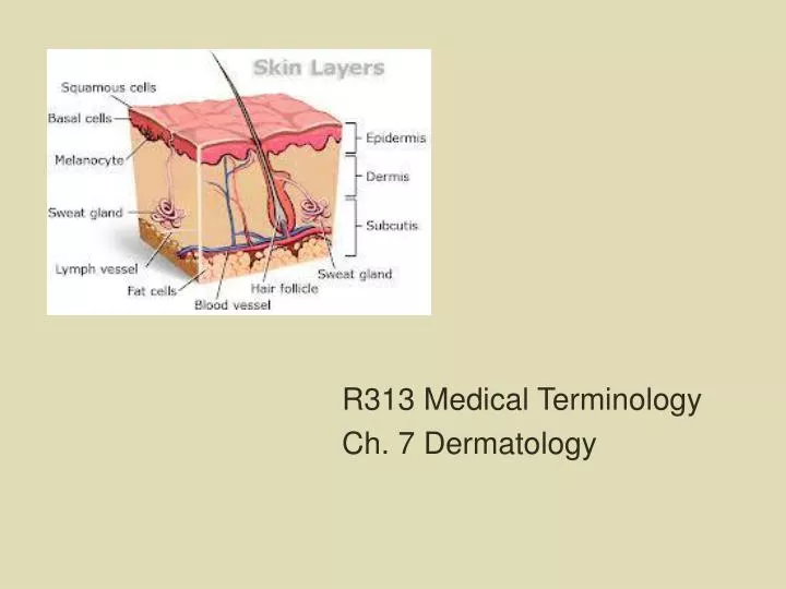
Anatomy and Physiology Integumentary System: skin (epidermis and dermis) sebaceous glands sweat glands hair nails Function: protects the body and is the first line of defense against invading microorganisms;the sense of touch.
Anatomy Skin layers: 1. Epidermis 2. Dermis 3. Subcutaneous
Characteristics of the Epidermis: -epithelial tissue -covers the external surface of the body and mucous membranes. -outer epidermis is the corneal layer. -lower epidermis is the basal layer. -external cells contain keratin, protein that gives skin a waterproofing ability. -cells also contain melanocytes that produce melanin, a protein pigment that gives skin its color; absorbs UV light from the sun. -dead cells are removed through a process called exfoliation.
Characteristics of the Dermis: -Connective Tissue -Thicker layer than the epidermis -Contains collagen fibers (firm, white protein) and elastin fibers (elastic, yellow protein). -Contains: blood vessles neurons (nerve cells) hair follicles sebaceous glands (oil glands; secrete sebum) sweat glands (sudoriferous glands; secrete water, sodium, waste products) -Has dermatome’s: specific area on the skin that sends sensory information to the spinal cord.
Hair -Covers most of the body. -Additional facial, axillary, and pubic hairs appear during puberty. -Contains melanocytes and keratin. -Forms in a hair follicle in the dermis. -Piloerection
Tran Van Hay Hair Length = 20’ 3.6” XieQiupingHair Length = 18’ 5.54”
Nails -Cover and protect the distal ends of the fingers and toes. -Each nail consists of a nail plate, nail bed, cuticle, lunula, and nail root.
Characteristics of the Subcutaneous Tissue -Loose, connective tissue directly beneath the dermis of the skin. -Composed of adipose tissue that contains lipocytes (fat-storing cells). -Provides a layer of insulation to conserve internal body heat. -Acts as a cushion to protect the bones and internal organs.
Physiology of an Allergic Reaction -An allergy or allergic reaction is a hypersensitivity response to certain types of antigens known as allergens. Allergens: foods pollens molds animal dander dust chemicals drugs
-The basis of all allergic reactions is the release of histamine from basophils in the blood and mast cells in the connective tissue. -A local reaction occurs when an allergen touches the skin or mucous membranes. -Anaphylaxis is a severe systemic allergic reaction that can be life-threatening. (anaphylactic shock) Epi-pen
Diseases & Conditions -Dermatitis -Edema -Hemorrhage petechiae contusion (bruise) ecchymosis (large contusion) hematoma
Neoplasm- benign or malignant growth occurring on the skin. Pruritus- itching Rash Wound Xeroderma- excessive dryness of the skin
Skin Color Conditions: Albinism- lack of pigment because of a genetic mutation. Cyanosis- bluish-purplish discoloration of the skin. Erythema- reddish discoloration of the skin. Jaundice- yellowing of the skin; liver cannot process bilirubin. Necrosis- blackish discoloration of the skin. Pallor- paleness due to lack of blood supply. Vitiligo- autoimmune disease where the melanocytes are destroyed causing white patches.
Figure 7-7 Necrosis and pallo Meyer/Custom Medical Stock Photo, Inc.
Skin Injuries: Abrasion Blister Callus (corn)
Burn Types 1st Degree Burns: -a burn that affects the epidermis only, causing erythema without blistering. 2nd Degree Burns: -a burn that affects the epidermis and the dermis, classified as superficial (involving the epidermis and the papillary dermis) or deep (extending into the reticular dermis). Called also partial thickness burn. 3rd Degree Burns: -a burn that destroys both the epidermis and the dermis, often also involving the subcutaneous tissue. Called also full-thickness burn 4th Degree Burns: -a burn that extends deeply into the subcutaneous tissue; it may involve muscle, fascia, or bone.
First Degree Burn Second Degree Burn
Third Degree Burn Fourth Degree Burn
Cicatrix- collagen scar tissue; keloid scar tissue formed from an overproduction of collagen. Decubitus Ulcer (bed sore)
Excoriation- scratch on the skin Laceration- deep wound
Skin Infections: Abscess- local pus containing pocket Cellulitis- inflammation and infection of the connective tissues. Herpes- caused by a virus Type 1- lips (cold sores) Type 2- HSV; sexually transmitted (genital herpes) Herpes Whitlow- infection at the base of the fingernail Herpes Varicella Roster- skin rash; chickenpox; adult version is called Shingles.
Figure 7-13 Shingles Gill/Custom Medical Stock Photo, Inc.
Tinea- fungal infection (ringworm, jock itch, athletes’s foot) Verruca (warts)- skin lesion caused by the human papillomavirus on the hands, fingers, or feet.
Skin Infestations: Pediculosis- lice Scabies- parasitic mites
Allergic Skin Conditions: Contact Dermatitis- reaction caused by an allergen coming in contact with the skin. Urticaria- hives, wheal & welts.
Benign Skin Markings and Neoplasms: Actinic Keratoses- raised, dry, rough areas of the skin. Freckle- grouping of melanocytes from sun exposure. Hemangioma- growth of superficial blood vessels.
Nevus- birth marks Papilloma- small protrusion of epidermis and dermis. Premalignant Skin Lesions- abnormal skin lesions that are not yet cancerous. Senile Lentigo- light to dark brown macules from exposure to the sun. (sun spots) Syndactyly- webbed fingers or toes; polydactyly is extra fingers or toes. Xanthoma- benign growth that is a yellow nodule or plaque on the hands, elbows, knees, or feet.
Malignant Neoplasms of the Skin: Cancer- malignant Basal Cell Carcinoma- most common type Malignant Melanoma- can spread to other parts of the body. A- asymmetry B- border C- color D- diameter
Squamous Cell Carcinoma Kaposi’s Sarcoma- skin cancer that begins in the connective tissue or lymph nodes. Common in AIDS patients.
Autoimmune Diseases with Skin Symptoms: Psoriasis- excessive number of epidermal cells; causes redness and itching; scales and plaque. Scleroderma- skin and internal organs progressively harden due to deposits of collagen. Systemic Lupus Erythematosus (SLE)- deterioration of the collagen in the skin and connective tissues; joint pain, sun sensitivity, fatigue.
Figure 7-22 Psoriasis NMSB/Custom Medical Stock Photo, Inc.
Diseases of the Sebaceous Glands: Acne Vulgaris- during puberty, sebaceous glands secrete large amounts of sebum. Acne Rosacea- middle-aged patients, sebaceous glands secrete large amounts of sebum, causes blotchy redness and edema. Seborrhea- overproduction of sebum at times other than puberty; also called cradle cap in infants and eczema in adults.
Diseases of the Sweat Glands: Anhidrosis- absence of sweat glands. Diaphoresis- profuse sweating.
Diseases of the Hair: Alopecia- chronic loss of scalp hair. Folliculitis- infection of the hair follicle; often occurs after shaving. Hirsutism- presence of excessive, dark hair on the forearms and over the lip of women. Pilonidal Sinus- abnormal passageway that begins as a large, abnormal hair follicle that contains a hair that is never shed.
Diseases of the Nails: Clubbing- abnormally curved fingernails and stunted growth of fingers. Onychomycosis- fungal infection of the fingernails or toenails. Paronychia- bacterial infection of the skin next to the cuticle.
Laboratory and Diagnostic Procedures: Allergy Skin Testing
Culture and Sensitivity (C & S)- test done on pus of a bacteria infection to determine course of treatment. RAST- blood test done to determine allergies. Skin Scraping- sampling of skin taken to be observed in determining if a patient has ringworm. TzanckTest- skin scraping done to test fluid for the presence of a virus. Wood’s Lamp- UV light used to highlight areas of the skin in a darkened room.
Medical and Surgical Procedures: Botox Injections- the drug Botox is injected into the skin to paralyze the muscle. Collagen Injections- collagen is a protein, helps to plump the skin to reduce wrinkles and scarring.
Cryosurgery- liquid nitrogen is put onto a wart or mole to remove it. Curettage- a tool called a curet is used to remove a superficial skin lesion. Debridement- procedure to remove necrotic tissue from a burn, wound, or ulcer. Electrosurgery- electricity is used to remove a nevus, wart, or small malignant lesion. Incision and Drainage- a scalpel is used to make a cut to drain infection from an abscess or cyst.
Laser Surgery- a laser is used to remove birthmarks, tattoos, enlarged blood vessels, or hair. Skin Examination- examining a patient’s skin. Skin Resurfacing- removal of acne scars, tattoo’s, wrinkles. Chemical Peel- use a chemical to remove the outer layers of the epidermis. Dermabrasion- removal of the outer epidermal layers using a spinning wire brush or diamond surface.
Laser Skin Resurfacing- use of a laser to remove outer layers of the epidermis and/or dermis. Microdermabrasion- skin resurfacing done using aluminum oxide crystals. Suturing- use of sutures to bring edges of skin together to allow for better healing.
Surgical Procedures: Biopsy- procedure done to remove a skin lesion for testing and diagnosis. Excisional Biopsy- use of a scalpel to remove a skin lesion. Incisional Biopsy- uses of a scalpel to remove part of a skin lesion. Needle Aspiration- use of a needle to remove cells for testing from a skin lesion.
Punch Biopsy- circular metal cutter is used to remove a plug shaped core of skin. Shave Biopsy- use of a razor blade to shave of a skin lesion. Dermatoplasty- any type of plastic surgery of the skin. Liposuction- produce used to remove excess adipose tissue.