Download
1 / 34
340 likes | 352 Views
Explore the role of Galectin-1 as a potential biomarker for predicting Sorafenib resistance in liver cancer using SILAC and iTRAQ quantitative proteomic analysis. Investigate EMT process in HuH-7/R cells and tumor xenograft models. Functions, pathways, and network analysis of differentially expressed proteins. Validation through Western blotting and ChIP assays.

E N D
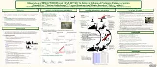
Integrated Stable Isotope Labeling by Amino Acids in Cell Culture (SILAC) and Isobaric Tags for Relative and Absolute Quantitation (iTRAQ) Quantitative Proteomic Analysis Identifies Galectin-1 as a Potential Biomarker for Predicting Sorafenib Resistance in Liver Cancer MOL CELL PROTEOMICS5.912 2017-05-25 IMI CONFIDENTIAL
Background 索拉非尼(Sorafenib)属于多激酶抑制剂,是一种新型多靶点抗肿瘤药物。具有抑制肿瘤细胞增殖和血管形成的双重作用,即一方面可以抑制信号传导系统Raf抑制肿瘤细胞增生;另一方面通过抑制血管内皮生长因子受体(vascular endothelial growth factor receptor, VEGFR)和血小板衍生生长因子受体(platelet-derived growth factor receptor, PDGFR)等酪氨酸激酶受体从而抑制肿瘤新生血管的形成和切断肿瘤细胞的营养供应而达到遏制肿瘤生长目的。
EMT的发生主要包括细胞形态学的改变、细胞间粘附结构的缺失和细胞免疫表型的变化:EMT的发生主要包括细胞形态学的改变、细胞间粘附结构的缺失和细胞免疫表型的变化: (1)细胞形态学上:上皮细胞极性缺失,细胞变为梭形; (2) 细胞间超微结构发生变化:骨架结构由以角蛋白为主的转化为以波形蛋白(Vimentin)为主,同时细胞间的连接结构蛋白如闭锁小带蛋白1(ZO-1)和泡膜蛋白1(VMP1)下调或缺失,导致细胞间粘附能力降低; (3) 细胞的免疫表型:上皮细胞标记物E-cadherin表达降低,-catenin向细胞核内转移,间质细胞标记物N-cadherin、Vimentin等表达上调,这一过程使细胞失去上皮表型,表现出间质细胞特性。
细胞缺氧模型建立: 1. 化学缺氧法:细胞培养基中加入化学诱导剂如二氯化钴(Cocl2)、氰化物、亚硫酸钠等进行化学诱导,这些化学试剂可造成细胞内呼吸链功能障碍或者导致培养基内氧浓度降低,从而使细胞出现缺氧。 2. 物理缺氧法:应用三气培养箱根据调控不同浓度的O2, CO2和N2使细胞出现不同程度的缺氧。 用三气培养箱、亚硫酸钠和Cocl2进行肿瘤细胞缺氧研究发现,三种方法都能导致细胞缺氧,引起细胞中HIF-1表达的升高,其中三气培养箱法和Cocl2诱导法还能使细胞发生EMT的改变,通过低氧浓度或Cocl2干预后,细胞中的EMT相关蛋白的表达发生了改变,细胞中HIF-1和Vimentin表达显著上调,而E-cadherin的表达下调,由此说明这两种方法均能引起细胞缺氧,并能致使细胞发生EMT。 Cocl2已被广泛用于细胞缺氧致EMT模型,该方法造模稳定。
Methods HuH-7:HCC cell HuH-7R:sorafenib-resistant HCC cell line Tumor xenograft model: HuH-7/HuH-7R:subcutaneously injecting (Tumor dimensions and tumor volume)OR tail vein inoculation (H&E) 91 advanced HCC patients/17 healthy volunteers SILAC (for in vitro labeling) iTRAQ (for in vivo labeling) Off-line 2D-LC-MS/MS Protein Identification and Quantification Mascot search engine (http://www.matrixscience.com) Ingenuity Pathway Analysis Tool (IPA)(http://www.ingenuity.com) Immunoblotting and Immunohistochemistry (IHC) Chromatin Immunoprecipitation (ChIP) Assays
1. Functional Analyses of HuH-7 and HuH-7RCells HuH-7R cells possess a more aggressive phenotype than HuH-7 cells
The sorafenibresistant cells showed an activation of the EMT process with enhanced invasive and metastatic potentials.
2. Identification and Quantification of Differentially Expressed Proteins in HuH-7 and HuH7R Cells and Cell-Derived Tumors
4616 2836 A total of 2,450 proteins common to both SILAC and iTRAQ experiments were reliably. 699 644 156 proteins were differentially expressed between HuH-7 and HuH-7Rcells. 81 proteins was increased in HuH-7R cells (>2.0-fold) 75proteins was decreased (<0.5-fold) 301 392 252 398
3. Biological Function, Pathway, and Network Analysis Red :EMT-related proteins 9 increased; 1 decreased 详见Table 1 和Table 2
81 up-regulated proteins • 75 down-regulated proteins
10 significantly differentially expressed proteins mainly participated in cellular movement or invasion (ANXA1, ANXA2, CCDC88A, CTGF, EPHA2, EZR, LGALS1(galectin-1), IQGAP1, RALGAPA2and vimentin).
4. Selected In Vitro- and In Vivo-Overexpressed Proteins Associated with Epithelial-Mesenchymal Transition (EMT) 6 proteins associated with EMT including vimentin, CTGF, IQGAP1, galectin-1, ezrin, and annexinA2 were selected. SILAC spectra are shown for sorafenib-regulated proteins
Validation of comparative proteomic results by western blotting
A total of 22 proteins were putative secreted proteins (SignalPprogram). Two of these candidates—galectin-1 (半乳糖凝集素1)and CTGF(结缔组织生长因子)—werehighly expressed in HuH-7R cells. Proteins dysregulated in HuH-7Rcells that used as HCC serum biomarkers for predicting sorafenibresistance?
5. Galectin-1 Knockdown Inhibits HuH-7R Cell Proliferation, Migration, and Invasion, and Restores SorafenibSensitivity
IC50 value HuH-7 cells (4.13) Knockdown of galectin-1 not only attenuates cell proliferation and metastasis in HuH-7R cells, it also restores sorafenibsensitivity.
6. High Expression of Galectin-1 in HuH-7R Cells Promotes Tumorigensis and Pulmonary Metastasis In Vivo Ki67作为标记细胞增殖状态的抗原,阳性说明癌细胞增殖活跃。
HuH-7R cells have greater tumorigenic and metastatic potential than HuH-7 cells in vivo.
7. Galectin-1 Expression is Regulated by PI3K/AKT, mTOR and HIF-1 Pathways Bioinformatics analyses indicated that up-regulation of the mTORsignaling pathway involved in facilitating the sorafenibresistance of HuH-7Rcells.
AKT and mTOR pathways are involved in galectin-1 up-regulation.
HIF-1作为肿瘤在低氧条件下最主要的调控因子,在促进肿瘤的侵袭转移过程中起着关键作用。在肿瘤缺氧微环境中, HIF-1能诱导和(或)调控EMT的过程。 Galectin-1 is a direct target of HIF-1. Cocl2(二氯化钴)缺氧诱导剂建立体外细胞缺氧模型。
The expression of galectin-1 is mediated by the PI3K/AKT/mTOR/HIF-1 pathway.
8. Prognostic Value of Galectin-1 in Advanced HCC Patients PFS(Progression-Free-Survival, 无进展生存期):从治疗开始至肿瘤客观进展或任何原因的死亡的时间。 OS(Overall survival, 总生存期):从治疗开始至随访截至时间所观察到的存活时间。
52 39 High galectin-1 serum level is associated with poor treatment efficacy of sorafenib, and shorter survivals in advanced HCC patients treated with sorafenib.
多因素分析显示表达galectin-1可作为肝癌无进展生存期和总生存期的独立预后因素。多因素分析显示表达galectin-1可作为肝癌无进展生存期和总生存期的独立预后因素。 综合表明过表达galectin-1导致sorafenib抵抗。