Download
1 / 47
470 likes | 492 Views
Protein Structures and Methods. Andy Howard Introductory Biochemistry 15 September 2014, IIT. Proteins are worth studying. We ’ ll offer an overview of what proteins look like Then we ’ ll perform a quick overview of methods of studying proteins Purification methods Analytical methods

E N D
Protein Structures and Methods Andy Howard Introductory Biochemistry15 September 2014, IIT
Proteins are worth studying • We’ll offer an overview of what proteins look like • Then we’ll perform a quick overview of methods of studying proteins • Purification methods • Analytical methods • Structural methods Protein Fundamentals
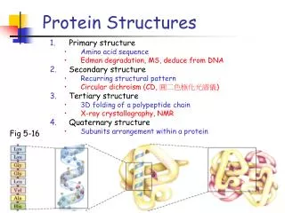
Levels of protein structure Primary Secondary Tertiary Quaternary Domains TIM Barrels Generalizations about structure Methods of purifying proteins Plans Protein Fundamentals
Levels of Protein Structure:G&G §5.1 • We conventionally describe proteins at four levels of structure, from most local to most global: • Primary: linear sequence of peptide units and covalent disulfide bonds • Secondary: main-chain H-bonds that define short-range order in structure • Tertiary: three-dimensional fold of a polypeptide • Quaternary: Folds of multiple polypeptide chains to form a complete oligomeric unit Protein Fundamentals
Components of secondary structure (G&G §6.3) • , 310, helices • pleated sheets and the strands that comprise them • Beta turns • More specialized structures like collagen helices Protein Fundamentals
An accounting for secondary structure: phospholipase A2 Protein Fundamentals
Alpha helix (G&G fig. 6.6) Protein Fundamentals
Characteristics of helices(G&G Fig. 6.9) • Hydrogen bonding from amino nitrogen to carbonyl oxygen in the residue 4 earlier in the chain • 3.6 residues per turn • Amino acid side chains face outward, for the most part • ~ 10 residues long in globular proteins Protein Fundamentals
What would disrupt this? • Not much: the side chains don’t bump into one another • Proline residue will disrupt it: • Main-chain N can’t H-bond • The ring forces a kink • Glycines sometimes disrupt because they tend to be flexible Protein Fundamentals
Other helices • NH to C=O four residues earlier is not the only pattern found in proteins • 310 helix is NH to C=O three residues earlier • More kinked; 3 residues per turn • Often one H-bond of this kind at N-terminal end of an otherwise -helix • helix: even rarer: NH to C=O five residues earlier Protein Fundamentals
Beta strands • Structures containing roughly extended polypeptide strands • Extended conformation stabilized by inter-strand main-chain hydrogen bonds • No defined interval in sequence number between amino acids involved in H-bond Protein Fundamentals
Sheets: roughly planar(G&G fig. 6.10) • Pleats straighten H-bonds • Side-chains roughly perpendicular from sheet plane • Consecutive side chains up, then down • Minimizes intra-chain collisions between bulky side chains Protein Fundamentals
Anti-parallel beta sheet • Neighboring strands extend in opposite directions • Complementary C=O…N bonds from top to bottom and bottom to top strand • Slightly pleated for optimal H-bond strength Protein Fundamentals
Parallel Beta Sheet • N-to-C directions are the same for both strands • You need to get from the C-end of one strand to the N-end of the other strand somehow • H-bonds at more of an angle relative to the approximate strand directions • Therefore: more pleated than anti-parallel sheet Protein Fundamentals
Beta turns • Abrupt change in direction • , angles arecharacteristic of beta • Main-chain H-bonds maintained almost all the way through the turn • Jane Richardson and others have characterized several types Protein Fundamentals
Collagen triple helix • Three left-handed helical strands interwoven with a specific hydrogen-bonding interaction • Every 3rd residue approaches other strands closely: so they’re glycines Protein Fundamentals
Hydrogen bonds, revisited • Protein settings, H-bonds are almost always: • Between carbonyl oxygen and hydroxyl:(C=O ••• H-O-) • between carbonyl oxygen and amine:(C=O ••• H-N-) • –OH to –OH, –OH to –NH, … less significant • These are stabilizing structures • Any stabilization is (on its own) entropically disfavored; • Sufficient enthalpic optimization overcomes that! • In general the optimization is ~ 3-7 kJ/mol Protein Fundamentals
Secondary structures in structural proteins • Structural proteins often have uniform secondary structures • Seeing instances of secondary structure provides a path toward understanding them in globular proteins • Examples: • Alpha-keratin (hair, wool, nails, …):-helical • Silk fibroin (guess) is -sheet Protein Fundamentals
Alpha-keratin • Actual -keratins sometimes contain helical globular domains surrounding a fibrous domain • Fibrous domain: long segments of regular -helical bonding patterns • Side chains stick out from the axis of the helix Protein Fundamentals
Silk fibroin • Antiparallel beta sheets running parallel to the silk fiber axis • Multiple repeats of (Gly-Ser-Gly-Ala-Gly-Ala)n Protein Fundamentals
Secondary structure in globular proteins • Segments with secondary structure are usually short: 2-30 residues • Some globular proteins are almost all helical, but even then there are bends between short helices • Other proteins: mostly beta • Others: regular alternation of , • Still others: irregular , , “coil” Protein Fundamentals
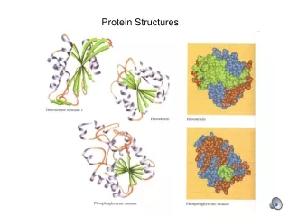
Tertiary Structure(G&G §6.4) • The overall 3-D arrangement of atoms in a single polypeptide chain • Made up of secondary-structure elements & locally unstructured strands • Described in terms of sequence, topology, overall fold, domains • Stabilized by van der Waals interactions, hydrogen bonds, disulfides, . . . Protein Fundamentals
Generalizations about Tertiary Structure • Most globular proteins contain substantial quantities of secondary structure • The non-secondary segments are usually short; few knots or twists • Most proteins fold into low-energy structures—either the lowest or at least in a significant local minimum of energy • Generally the solvent-accessible surface area of a correctly folded protein is small Protein Fundamentals
Hydrophobic in, -philic out • Aqueous proteins arrange themselves so that polar groups are solvent-accessible and apolar groups are not • The energetics of protein folding are strongly driven by this hydrophobic in, hydrophilic out effect • Exceptions are membrane proteins Protein Fundamentals
Quaternary structure • Arrangement of individual polypeptide chains to form a complete oligomeric, functional protein • Individual chains can be identical or different • If they’re the same, they can be coded for by the same gene • If they’re different, you need more than one gene Protein Fundamentals
Generalizations about quaternary structure • Considerable symmetry in many quaternary structure patterns(see G&G section 6.5) • Weak polar and solvent-exclusion forces add up to provide driving force for association • Many quaternary structures are necessary to function:often the monomer can’t do it on its own Protein Fundamentals
Not all proteins have all four levels of structure • Monomeric proteins don’t have quaternary structure • Tertiary structure: subsumed into 2ndry structure for many structural proteins (keratin, silk fibroin, …) • Some proteins (usually small ones) have no definite secondary or tertiary structure; they flop around! Protein Fundamentals
Protein Topology • Description of the connectivity of segments of secondary structure and how they do or don’t cross over Protein Fundamentals
TIM barrel • Alternating , creates parallel -pleated sheet • Bends around as it goes to create barrel Protein Fundamentals
How do we visualize protein structures? • It’s often as important to decide what to omit as it is to decide what to include • Any segment larger than about 10Å needs to be simplified if you want to understand it • What you omit depends on what you want to emphasize Protein Fundamentals
Styles of protein depiction • All atoms • All non-H atoms • Main-chain (backbone) only • One dot per residue (typically at C) • Ribbon diagrams: • Helical ribbon for helix • Flat ribbon for strand • Thin string for coil Protein Fundamentals
How do we show 3-D? • Stereo pairs • Rely on the way the brain processes left- and right-eye images • If we allow our eyes to go slightly wall-eyed or crossed, the image appears three-dimensional • Dynamics: rotation of flat image • Perspective (hooray, Renaissance) Protein Fundamentals
Green fluorescent protein Yang et al. (1999) Nature Biotechnology 14:1246 Protein Fundamentals
A little more complex Endonuclease V (Dalhus et al (2009) NSMB 16:138) Protein Fundamentals
Stereo pair: Release factor 2/3Klaholz et al, Nature (2004) 427:862 Protein Fundamentals
A little more complex: AligningCytochrome C5 & Cytochrome C550 Protein Fundamentals
Mixed:hen egg-white lysozyme PDB 2vb10.65Å, 14.2kDa Mostly helical:E.coli RecG – DNAPDB 1gm53.24Å, 105 kDa Ribbon diagrams Protein Fundamentals
The Protein Data Bank • http://www.rcsb.org/ • This is an electronic repository for three-dimensional structural information of polypeptides and polynucleotides • 103015 structures as of September 2013 • Most are determined by X-ray crystallography • Smaller number are high-field NMR structures • A few calculated structures, most of which are either close relatives of experimental structures or else they’re small, all-α-proteins Protein Fundamentals
What you can do with the PDB • Display structures • Look up specific coordinates • Run clever software that compares and synthesizes the knowledge contained there • Use it as a source for determining additional structures Protein Fundamentals
Domains • Proteins (including single-polypeptide proteins) often contain roughly self-contained domains • Domains often separated by linkers • Linkers sometimes flexible or extended or both • Cf. fig. 6.38 in G&G Protein Fundamentals
Protein Purification • Why do we purify proteins? • To get a basic idea of function we need to see a protein in isolation from its environment • That necessitates purification • An instance of reductionist science • Full characterization requires a knowledge of the protein’s action in context Protein Fundamentals
Salting Out • Most proteins are less soluble in high salt than in low salt • In high salt, water molecules are too busy interacting with the primary solute (salt) to pay much attention to the secondary solute (protein) • Various proteins differ in the degree to which their solubility disappears as [salt] goes up • We can separate proteins by their differential solubility in high salt. Protein solubility, mg/ML 0 2 [Salt], M Protein Fundamentals
How to do it • Dissolve protein mixture in highly soluble salt like Li2SO4, (NH4)2SO4, NaCl • Increase [salt] until some proteins precipitate and others don’t • You may be able to recover both: • The supernatant (get rid of salt; move on) • The pellet (redissolve, desalt, move on) • Typical salt concentrations > 1M Protein Fundamentals
Dialysis • Some plastics allow molecules to pass through if and only ifMW < Cutoff • Protein will stayinside bag, smaller proteins will leave • Non-protein impurities may leave too. Protein Fundamentals
Gel-filtration chromatography • Pass a protein solution through a bead-containing medium at low pressure • Beads retard small molecules • Beads don’t retard bigger molecules • Can be used to separate proteins of significantly different sizes • Suitable for preparative work Protein Fundamentals
Ion-exchange chromatography • Charged species affixed to column • Phosphonates (-) retard (+)charged proteins:Cation exchange • Quaternary ammonium salts (+) retard (-)charged proteins:Anion exchange • Separations facilitated by adjusting pH Protein Fundamentals
Affinity chromatography • Stationary phase contains a species that has specific favorable interaction with the protein we want • DNA-binding protein specific to AGCATGCT: bind AGCATGCT to a column, and the protein we want will stick; every other protein falls through • Often used to purify antibodies by binding the antigen to the column Protein Fundamentals