Download

1 / 94
940 likes | 953 Views
Discover the intricate functions of the lymphatic system, how it maintains fluid balance, fights infections, and absorbs nutrients. Explore lymphatic vessels, nodes, organs, and cell types that play crucial roles in immunity. Learn about lymph flow mechanisms and the importance of lymphatic drainage.

E N D
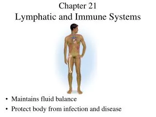
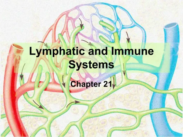
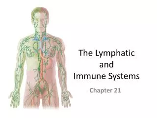
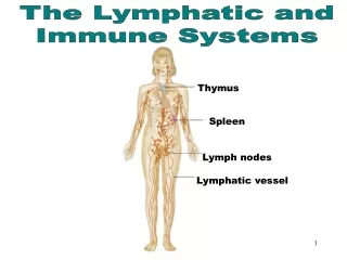
Chapter 21: Lymphatic and Immune Systems
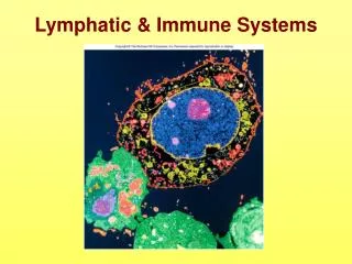
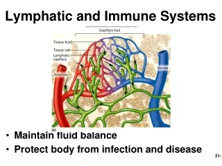
Lymphatic and Immune Systems • Maintain fluid balance • Protect body from infection and disease
Functions of Lymphatic System • Immunity • fluids from all capillary beds are filtered • immune cells stand ready to respond to foreign cells or chemicals encountered • Lipid absorption • Lacteals in small intestine absorb dietary lipids • Fluid recovery • absorbs plasma proteins and fluid (2 to 4 L/day) from tissues and returns it to the bloodstream • interference with lymphatic drainage leads to severe edema
Lymph and Lymphatic Capillaries • Lymph • clear, colorless fluid, similar to plasma but much less protein • Lymphatic capillaries • closed at one end • tethered to surrounding tissue by protein filaments • endothelial cells loosely overlapped • allow bacteria and cells entrance to lymphatic capillary • creates valve-like flaps that open when interstitial fluid pressure is high, and close when it is low
Lymphatic Vessels • Larger ones composed of 3 layers • tunica interna: endothelium and valves • tunica media: elastic fibers, smooth muscle • tunica externa: thin outer layer
Route of Lymph Flow • Lymphatic capillaries • Collecting vessels: course through many lymph nodes • Lymphatic trunks: drain major portions of body • Collecting ducts : • right lymphatic duct – receives lymph from R arm, R side of head and thorax; empties into R subclavian vein • thoracic duct - larger and longer, begins as a prominent sac in abdomen called the cisterna chyli; receives lymph from below diaphragm, L arm, L side of head, neck and thorax; empties into L subclavian vein
Mechanisms of Lymph Flow • Lymph flows at low pressure and speed • Moved along by rhythmic contractions of lymphatic vessels • stretching of vessels stimulates contraction • Flow aided by skeletal muscle pump • Thoracic pump aids flow from abdominal to thoracic cavity • Valves prevent backward flow • Rapidly flowing blood in subclavian veins, draws lymph into it • Exercise significantly increases lymphatic return
Lymphatic Cells • Natural killer (NK) cells • responsible for immune surveillance • T lymphocytes • mature in thymus • B lymphocytes • activation causes proliferation and differentiation into plasma cells that produce antibodies • Antigen Presenting Cells • macrophages (from monocytes) • dendritic cells (in epidermis, mucous membranes and lymphatic organs) • reticular cells (also contribute to stroma of lymph organs)
Lymphatic Tissue • Diffuse lymphatic tissue • lymphocytes in mucous membranes and CT of many organs • Mucosa-Associated Lymphatic Tissue (MALT): prevalent in passages open to exterior • Lymphatic nodules • dense oval masses of lymphocytes, congregate in response to pathogens • Peyer patches: more permanent congregation, clusters found at junction of small to large intestine
Lymphatic Organs • At well defined sites; have CT capsules • Primary lymphatic organs • site where T and B cells become immunocompetent • red bone marrow and thymus • Secondary lymphatic organs • immunocompetent cells populate these tissues • lymph nodes, tonsils, and spleen
Lymph Node • Lymph nodes - only organs that filter lymph • Fewer efferent vessels, slows flow through node • Capsule gives off trabeculae, divides node into compartments containing stroma (reticular CT) and parenchyma (lymphocytes and APCs) subdivided into cortex (lymphatic nodules) and medulla • reticular cells, macrophages phagocytize foreign matter • lymphocytes respond to antigens • lymphatic nodules-germinal centers for B cell activation
Lymphadenopathy • Collective term for all lymph node diseases • Lymphadenitis • swollen, painful node responding to foreign antigen • Lymph nodes are common sites for metastatic cancer • swollen, firm and usually painless
Lymph Node Fig. 21.12 a and b
Tonsil • Covered by epithelium • Pathogens get into tonsillar crypts and encounter lymphocytes
Location of Tonsils • Palatine tonsils • pair at posterior margin of oral cavity • most often infected • Lingual tonsils • pair at root of tongue • Pharyngeal tonsil (adenoid) • single tonsil on wall of pharynx
Thymus • Capsule gives off trabeculae, divides parenchyma into lobules of cortex and medulla • Reticular epithelial cells • form blood thymus barrier in cortex • isolates developing T lymphocytes from foreign antigens • secretes hormones (thymopoietin, thymulin and thymosins) • to promote development and action of T lymphocytes • Very large in fetus; after age 14 begins involution • in elderly mostly fatty and fibrous tissue
Spleen • Parenchyma appears in fresh specimens as • red pulp: sinuses filled with erythrocytes • white pulp: lymphocytes, macrophages; surrounds small branches of splenic artery • Functions • blood production in fetus • blood reservoir • RBC disposal • immune reactions: filters blood, quick to detect antigens
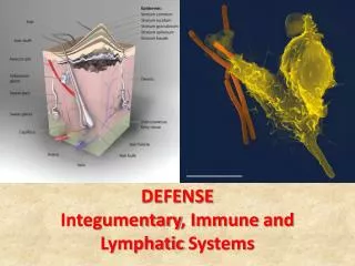
Defenses Against Pathogens • Nonspecific defenses - broadly effective, no prior exposure • first line of defense • external barriers • second line of defense • phagocytic cells, antimicrobial proteins, inflammation and fever • Specific defense - results from prior exposure, protects against only a particular pathogen • third line of defense • immune system
External Barriers • Skin • toughness of keratin • dry and nutrient-poor • defensins: peptides, from neutrophils attack microbes • lactic acid (acid mantle) is a component of perspiration • Mucous membranes • stickiness of mucus • lysozyme: enzyme destroys bacterial cell walls • Subepithelial areolar tissue • tissue gel: viscous barrier of hyaluronic acid • hyaluronidase: enzyme used by pathogens to spread
Leukocytes and Cutaneous Defenses • Neutrophils • Eosinophils • Basophils • Monocytes • Lymphocytes
Neutrophils • Phagocytize bacteria • Create a killing zone • degranulation • lysosomes discharge into tissue fluid • respiratory burst • toxic chemicals are created (O2.-, H2O2, HClO)
Eosinophils • Phagocytize antigen-antibody complexes • Antiparasitic effects • Promote action of basophils, mast cells • Enzymes block excess inflammation, limit action of histamine
Basophils • Aid mobility and action of WBC’s by release of • histamine (vasodilator) • blood flow to infected tissue • heparin (anticoagulant) • prevents immobilization of phagocytes
Monocytes • Circulating precursors to macrophages • Specialized macrophages found in specific localities • dendritic cells • epidermis, oral mucosa, esophagus, vagina, and lymphatic organs • microglia (CNS) • alveolar macrophages (lungs) • hepatic macrophages (liver)
Lymphocytes • Circulating blood contains • 80% T cells • 15% B cells • 5% NK cells
Antimicrobial Proteins • Interferons • Complement system
Interferons • Secreted by certain cells invaded by viruses • generalized protection • diffuse to neighboring cells and stimulate them to produce antiviral proteins • activate natural killer cells and macrophages • destroy infected host cells • stimulate destruction of cancer cells
Complement System • Complement (C) proteins in blood that must be activated by pathogens • Pathways of complement activation: C3 split into C3a and C3b • classical pathway • requires antibody; specific immunity • alternate pathway • nonspecific immunity • lectin pathway • nonspecific immunity
Complement System • Mechanisms of action • enhanced inflammation • phagocytosis • promoted by opsonization • cytolysis • membrane attack complex forms on target cell • immune clearance • RBCs carry Ag-Ab complexes to macrophages in liver and spleen
Complement Activation Fig. 21.15
Membrane Attack Complex • Complement proteins form ring in plasma membrane of target cell causing cytolysis
Immune Surveillance • NK cells • destroy bacteria, transplanted cells, cells infected by viruses, and cancer cells • release perforins and granzymes
Action of NK cell Fig. 21.17
Inflammation • Defensive response to tissue injury • limits spread of pathogens, then destroys them • removes debris • initiates tissue repair • Cytokines • small proteins regulate inflammation and immunity; include • interferons, interleukins, tumor necrosis factor, and chemotactic factors
Inflammation • Suffix -itis denotes inflammation of specific organs • Cardinal signs • redness (erythema) caused by hyperemia ( blood flow) • swelling (edema) caused by capillary permeability and filtration • heat caused by hyperemia • pain caused by inflammatory chemicals (bradykinin, prostaglandins) secreted by damaged cells, pressure on nerves
Inflammation • Three major processes • mobilization of body defenses • containment and destruction of pathogens • tissue clean-up and repair
Mobilization of Defenses • Kinins, histamine, and leukotrienes are secreted by damaged cells, basophils and mast cells • stimulates vasodilation that leads to hyperemia • causes redness and heat • local metabolic rate, promotes cell multiplication and healing • dilutes toxins, provides O2, nutrients, waste removal • stimulates permeability of blood capillaries • allows blood cells, plasma proteins (antibodies, complement proteins, fibrinogen) into tissue • clotting sequesters bacteria, forms scaffold for tissue repair
Mobilization of Defenses • Leukocyte Deployment • margination • selectins cause leukocytes to adhere to blood vessel walls • diapedesis (emigration) • leukocytes squeeze between endothelial cells into tissue space
Containment and Destruction of Pathogens • Fibrinogen now in tissue clots, trapping pathogens • Heparin prevents clotting at site of injury • pathogens are in a fluid pocket surrounded by clot • Chemotaxis • leukocytes are attracted to chemotactic chemicals • Neutrophils are quickest to respond • phagocytosis • respiratory burst • secrete cytokines for recruitment of macrophages and neutrophils • macrophages and T cells secrete colony-stimulating factor to stimulate leukopoiesis
Tissue Cleanup • Monocytes the primary agents of cleanup arrive in 8 to 12 hours, become macrophages, • Edema venous flow, lymphatic flow that favors removal of bacteria and debris • Formation of pus • mixture of tissue fluid, cellular debris, dying neutrophils and microbes
Tissue Repair • Blood platelets and endothelial cells in injured area secrete a cytokine, PDGF, that stimulates fibroblasts to multiply and synthesize collagen • Facilitated by hyperemia that provides materials needed and heat that increases metabolism • Fibrin clot may provide a scaffold for repair • Pain limits use of body part allowing for repair
Fever • Defense mechanism: does more good than harm • promotes interferon activity • accelerating metabolic rate and tissue repair • inhibiting pathogen reproduction • A cytokine, interleukin 1, called a pyrogen • secreted by macrophages, stimulates anterior hypothalamus to secrete PGE which resets body thermostat higher > 105F may cause delirium, 111F- 115F, coma-death • Stages of fever • onset, stadium, defervescence