Download
1 / 39
400 likes | 783 Views
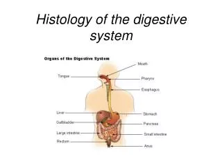
Digestive System Histology and Models. Glands and Tonsils on the Models. Submandibular gland Sublingual gland Parotid gland Palatine tonsil Pharyngeal tonsil. Glands. Parotid gland Submandibular. Glands. Sublingual Submandibular. Pharyngeal tonsil. Palatine tonsil. Stomach.

E N D
Glands and Tonsils on the Models • Submandibular gland • Sublingual gland • Parotid gland • Palatine tonsil • Pharyngeal tonsil
Glands • Parotid gland • Submandibular
Glands • Sublingual • Submandibular
Pharyngeal tonsil Palatine tonsil
Stomach Cardiac region Fundus region Lesser curvature Body region Greater curvature Pyloric region
Stomach Pyloric sphincter Plicae circularis Rugae Duodenum
PANCREAS AND SPLEEN MODELS • Kidney • Gall bladder • Pancreas • spleen (function is to remove foreign antigens and aging RBC’s) • duodenum • plicae circularis
Spleen Plicae circularis Pancreas Duodenum
Pancreas Gall bladder Spleen Kidney Plicae circularis Duodenum
GI MODEL • Stomach • Pancreas • Spleen • Pyloric sphincter • circular folds (known as plicae circularis; these are macroscopic folds in the mucosa) • ascending colon • transverse colon • descending colon • tenia coli • Jejunum (middle part of small intestine) • Ileum • Cecum • Ileocecal valve • Appendix • Rectum • NOTE: Plicae circularis, microvilli, and villi all increase the surface area of the small intestinal lining
Pyloric sphincter Stomach Duodenum Spleen Pancreas Plicae circularis
Transverse colon Tenia coli Ascending colon Descending colon Jejunum Rectum
Ileocecal valve Ileum Cecum Plicae circularis Appendix Tenia coli Rectum
INTESTINE MODEL • Stomach • Pancreas • Spleen • Pyloric sphincter • circular folds (known as plicae circularis; these are macroscopic folds in the mucosa) • ascending colon • transverse colon • descending colon • tenia coli • Jejunum (middle part of small intestine) • Ileum • Cecum • Ileocecal valve • Appendix • Rectum • NOTE: Plicae circularis, microvilli, and villi all increase the surface area of the small intestinal lining
Duodenum Pancreas Greater Omentum Tenia coli Transverse colon Jejunum Cecum
Pancreas Common bile duct Duodenum Tenia coli Descending colon Mesentery Appendix Ascending colon Cecum Sigmoid colon (first half)
Liver Models • gall bladder • cystic duct • common bile duct • falciform ligament
Liver CYSTIC DUCT, GALL BLADDER COMMON BILE DUCT (has been cut) Falciform ligament
SLIDES • Salivary gland • Tongue • taste bud • Small Intestine • Villi • Intestinal crypt
Tongue Taste buds
SLIDES • Small intestine • Villi, microvilli, and plicae circularis: (Serve to increase the surface area of the small intestine) • Intestinal crypts
Small Intestine Villus Intestinal crypt
SLIDES • Liver: the functional unit of the liver is the lobule. • central vein
Liver Lobule • Central vein
SLIDES • Spleen: removes foreign antigens and aging RBC's • Red pulp • White pulp • Pancreas • Islets • Acini • Be able to tell the difference between liver, spleen, pancreas, and kidney slides!
Spleen Red Pulp White pulp
Pancreas Islet of Langerhans (secretes insulin)
Pancreas Acinar cells (secrete enzymes) Islet of Langerhans (secretes insulin)
Kidney Tubule Glomerulus
Spleen Liver Central vein Red Pulp White pulp