Download
1 / 68
700 likes | 941 Views
Muscle: Contractions and Neural Control. More than ___ muscles in the animal body Characteristics of muscle dictate its function Mammalian and avian – long, unbranched, and threadlike that taper at both ends Muscle Fiber = ___________________. Function. Movement of skeleton

E N D
More than ___ muscles in the animal body Characteristics of muscle dictate its function Mammalian and avian – long, unbranched, and threadlike that taper at both ends Muscle Fiber = ___________________
Function • Movement of skeleton • Blood pressure/supply • Transport of ingesta • Generation of Body heat • Circulation of blood
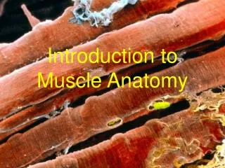
Muscle general anatomy • Tendon (tough connective tissue) • Muscle • Tendon
Embryonic development - Myoblasts • Muscle: Myotome (myoblast) cells migrate to various places in the embryo • Chemotaxis – ____________________ • Morphogens – chemicals produced in one area which effect distant cells
Growth • Hypertrophy: cells increase in _____ • Hyperplasia: addition of more cells • Growth: increase in muscle and bone • Fattening: accumulation of fat
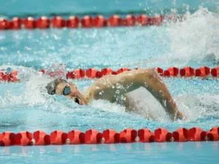
Skeletal Muscle Types: Slow- and Fast-Twitch Fibers • Divided on basis of contraction speed: • Slow-twitch (type I fibers). • ______________________: • Muscles which get used a lot, for long periods of time • Fowl that fly often, have dark breast (pectoral) meat • Fast-twitch (type II fibers). • _________________: fast twitch, easy to fatigue, lots of ___________________ present • ________________________: light colored pectorals • Intermediate: have a combination of the two • Most muscles • Differences due to different myosin ATPase isoenzymes that are slow or fast.
Upper Motor Neuron Control of Skeletal Muscles • Cerebellum: • Receives sensory input from muscle spindles • No descending tracts from the cerebellum. • Influences motor activity indirectly. • All output from cerebellum is inhibitory. • Aids motor coordination.
Upper Motor Neuron Control of Skeletal Muscles (continued) • Basal nuclei: • Profound inhibitory effects on the activity of lower motor neurons. • Damage to basal nuclei result in increased muscle tone.
Anatomy of Skeletal MuscleFigure 12.1 • Epimysium – CT that surrounds ______________ • Perimysium – CT that surrounds muscle bundle • Endomysium – CT that surrounds ____________________ • Sarcolemma – “____________” • Elastic CT that surrounds the myofibrils • Invaginations form a network of tubules called the transverse tubules “T-tubules”
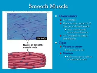
Muscle types • Striated muscle • ___________ • Autonomic control • Smooth muscle • _________________________ • No striations • Cardiac muscle • ____________________ • ______________________ control
Skeletal Muscle • Sarcoplasm – cytoplasm • Nuclei – multinucleated • Myofibrils – unique to muscle tissue • Long, thin, cylindrical rods 1 to 2 µm in diameter • Bathed in sarcoplasm • Consists of: ________________________ • Thick & Thin
Skeletal Muscle • Myofilaments • Thick are aligned parallel to each other • Thin , parallel to thick • These “bands” of parallel fibers give SM striated appearance • A and I bands • Called this due to light refractions • _________band is denser than___________ • Shows up darker in pictures • Both are bisected by thin, dense lines
Skeletal Muscle • A and I bands • I band is bisected by line called: _______ • Unit of myofibril between 2 different Z disks is called: ____________________ • Includes the A band and parts of the I band • Is the repeating structural unit of the myofibril • Basic unit in _____________ & ________________ • Length of sarcomere is not constant and will differ depending on relaxation or contraction
Myofilaments • Thick and thin differ in dimensions and chemical composition • Thick • 14 to 16 nanometers (nm = 1 billionth of a meter) • Constitute the ______________ of sarcomere • ____________________ is the predominant protein • held in position by other proteins some of which are located in the M line
Myofilaments • Thin • 6-8 nm in diameter • Extend 1.0 µm on either side of the Z disk • Filaments constitute the ________ of the sarcomere • Extend into the _________________ • I band only contains the ________ filaments
Myofilaments • ________________ – only thick filaments are present • ________________ – thick and thin are located • Shows 6 thin filaments surrounding each thick
Z- Disk • Z disk • Comprised of z-filaments which connect actin molecules • Z line is composed of 4 z-filaments which attach actin molecules from each sarcomere
Proteins of myfofibril • 20 different proteins associated with myofibrils • 6 proteins account for 90% of total myofibrillar protein • Decreasing order of abundance: • Myosin • Actin • Titin • Tropomyosin • Troponin • Nebulin
Proteins of myofibril • Major Contractile Proteins • Actin – 20% of myofibrillar protein Globular – G - actin Form a “super helix” Globular – G - actin Form a “super helix”
Actin myofilaments are made of globular actin (G-actin) monomers and other proteins.
Proteins of myofibril • Major Contractile Proteins • Myosin – 45% of myofibril protein Elongated Rod Thickened end: Head – 2 of them Thin end: Tail In-between: neck
Proteins of myofibril • Major Contractile Proteins Myosin • 2 fractions: ________ and __________meromyosin • Center of A band – myosin contains only rods, no heads • Known as pseudo H zone • Heads are functionally active during contraction • Form a cross-bridge with ______________
Proteins of myofibril • Tropomyosin 5% of myofibrillar protein • Lies in close contact with ____________ • Lies in grove with __________
Proteins of myofibril • Troponin: 5% of myofibril protein • In groove of _________________ • Lies astride the tropomyosin strands
Proteins of myofibril • Titin: 10% of myofibrillar protein • Scaffold for alignment of filaments during myofibril and sarcomere formation
Proteins of myofibril • Nebulin: 4% of myofibril protein • Anchors thin filaments to Z disks • Serves as a template for assembly / scaffold for stability of thin filaments
Muscle Contraction • Each myofibril contains myofilaments. • 2 major ones involved with contraction • Thick filaments aka ________________: • A bands contain thick filaments (primarily composed of myosin). • Myosin initiates the contraction • There an enzyme that converts ATP to ADP and Phosphate • Thin filaments aka ___________________: • I bands contain thin filaments (primarily composed of actin). • Most abundant protein • Major constituent of muscle • Center of each I band is Z disc.
Mechanisms of Contraction (continued) • Sarcomere: • _____ disc to ________ disc. • M lines: • Produced by protein filaments in a sarcomere. • Anchor myosin during contraction. • Titin: • Elastic protein that runs through the myosin from ____ line to ____________. • Contributes to elastic recoil of muscle.
Mechanisms of Contraction (continued) • Tropomyosin: • Part of the “___________” filament • Continuous stand that sits on actin • Exposes the actin binding site • Troponin: • Inhibits actin-activated myosin ATPase Activity
Sliding Filament Theory of Contraction • Sliding of filaments is produced by the actions of cross bridges. • Cross bridges are part of the myosin proteins that extend out toward actin. • Form arms that terminate in heads. • Each myosin head contains an ATP-binding site. • The myosin head functions as a myosin ATPase.
Sliding Filament Theory of Contraction (continued) • Muscle contracts: • Occurs because of sliding of thin filaments over and between thick filaments towards center. • Shortening the distance from Z disc to Z disc. • A bands: • Contain ______________. • Move closer together. • Do not shorten.
Sliding Filament Theory of Contraction (continued) • I bands: • Distance between A bands of successive ______________________. • Decrease in length. • H bands shorten. • Contain only _____________. • Shorten during contraction.
Contraction • Myosin binding site splits ATP to ADP and Pi. • ADP and Pi remain bound to myosin until myosin heads attach to actin. • Pi is released, causing the power stroke to occur. • Power stroke pulls actin toward the center of the A band. • ADP is released, when myosin binds to a fresh ATP at the end of the power stroke.
Contraction (continued) • Release of ADP upon binding to another _______, causes the cross bridge bond to break. • Cross bridges detach, ready to bind again. • Synchronous action: • Only 50% of the cross bridges are attached at any given time.
Regulation of Contraction • Regulation of cross bridge attachment to actin due to: • Tropomyosin:. • Lies within grove between double row of G-actin. • Troponin: • Attached to tropomyosin. • Serves as a switch for muscle contraction and relaxation. • In relaxed muscle: • Tropomyosin blocks binding sites on actin.
Role of Ca2+ in Muscle Contraction • Muscle Relaxation: • [Ca2+] in sarcoplasm low when tropomyosin blocks attachment. • Prevents muscle contraction. • Ca2+ is pumped back into the SR in the ____________________________. • Muscle relaxes.
Sarcoplasmic Reticulum & T Tubules • Membranous system of tubules and cisternae (reservoirs for calcium) • Found around each _________________ • T-tubules: • Associated with __________________ • T tubule runs transversely across the sarcomere at the AI junction.