Download
1 / 24
530 likes | 2.03k Views
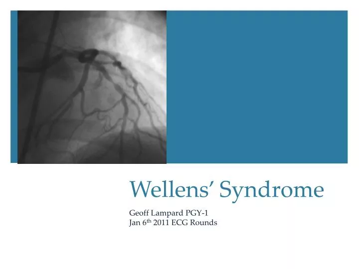
Wellens ’ Syndrome. Geoff Lampard PGY-1 Jan 6 th 2011 ECG Rounds. Case 1. 5 5 yo male with history of stable angina P resents to ED with ischemic chest pain. …. pain resolves with NTG…. Serum TnT returns normal What do you see in the ECG?. Case 2.

E N D
Wellens’ Syndrome Geoff Lampard PGY-1 Jan 6th 2011 ECG Rounds
Case 1 • 55yo male with history of stable angina • Presents to ED with ischemic chest pain
…. pain resolves with NTG….. • Serum TnT returns normal • What do you see in the ECG?
Case 2 • 57 yo male with 4/10 pressure-like chest pain • ASA, O2, 2 x SL NTG sprays with EMS • Pain now improving
Case 3 • 54M with pressure like RSCP, diaphoresis • NTG and morphine relieves pain. ECG follows:
Now 1 minute into the stress test…… • Anterior wall STEMI. • Immediately taken for cath and found an extensive proximal LAD lesion
Wellens’ Syndrome First recognised in early 1980’s in a subgroup of UA patients with precordial T-wave changes in the pain free periodwho developed large anterior MI’s. All patients who met their criteria had ≥ 50% LAD stenosis 26/145 patients admitted for UA developed characteristic ECG findings at or within 24hrs of admission
Key concept #1 Wellensis relatively common (14-18% of UA patients)
½ way through study, medical management was aborted and all were given preferential angiography 75% of medically managed patients developed anterior MI’s within 3 weeks
Key concept #2 Wellens is a preinfarction stage of CAD Mean time to infarction: 8.5 days
Criteria ECG findings: Symmetric, deeply inverted T waves in V2-3. Occasionally in V1,V4-6, or Biphasic T waves in V2-3plus Isoelectric or minimally elevated (<1mm) ST No precordial Q-waves History of angina Pattern in pain free state Normal or slightly elevated serum markers
Key concept #3 It is best seen during the pain-free period…. Get serial or pain-free ECG’s from UA patients!
Deep Inverted (figures A-C) ~75% of Wellens Biphasic pattern (D-F) ~25% of cases
But remember, the differential for t-wave inversion is large. Acute ischemia LVH BBB Late pericarditis PE CNS pathology Myocarditis Digitalis Old MI etc. etc. etc.
Key concept #4 Patients need early angiography. Get cardiology involved in the ED!
Wellens’ is not uncommon. Remember the 2 characteristic ECG changes Get a pain-free ECG! Wellens’ is a sign of impending MI. Requires urgent assessment.
De Zwann C, Bar FW, Wellens JHH: Characteristic electrocardiographic pattern indicating a critical stenosis high in left anterior descending coronary artery in patients admitted because of impending myocardial infarction. Am Heart J 1982. 103:730-736 De ZwannC, Bar FW, Janssen JH, et al: Angiographic and clinical characteristics of patents with unstable angina showing an ECG pattern indicating critical narrowing of the proximal LAD coronary artery. Am Heart J 1989. 117:657-665 RhinehardtJ, Brady WJ, Perron AD, Mattu A. Electrocardiographic Manifestations of Wellens’ Syndrome. Am Journal Emerg Med2002. 20:638-643. Lilaonitkul M, Ronbinson K, Roberts M. Wellens’ Syndrome: significance of ECG pattern recognition in the emergency department. Emerg Med J. 2009. 26:750-751 Questions?