Download
1 / 34
530 likes | 3.83k Views
The Management of SMA Syndrome. Dr Chun-fai LAU United Christian Hospital Joint Hospital Surgical Grand Round 11 Feb 2012. Case presentation. Mr. Leung M/63 Diagnosed to have localized CA sigmoid colon in Oct 2011 PMH Old CVA Ankylosing spondylitis. Case presentation.

E N D
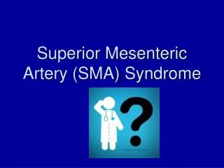
The Management of SMA Syndrome Dr Chun-fai LAU United Christian Hospital Joint Hospital Surgical Grand Round 11 Feb 2012
Case presentation Mr. Leung M/63 Diagnosed to have localized CA sigmoid colon in Oct 2011 PMH Old CVA Ankylosing spondylitis
Case presentation Lap converted open sigmoidectomy on 13 Oct 2011 Post-op required ICU care with ventilatory support for aspiration pneumonia Unable to tolerate oral feeding with repeated vomiting
Case presentation Postop CT image Dilatation of duodenum down to D3
Case presentation Postop CT image
Case presentation Decreased aortomesenteric distance 5.5mm Dilated duodenum down to D3 Suggestive of SMA syndrome
Case presentation Endoscopic and fluoroscopic guided feeding tube insertion on 8 Nov 2011 ( 3 week postop ) Failed conservative management Laparotomy with division of ligament of Treitz on 13 Dec 2011 ( 8 weeks postop ) Post-op Water soluble contrast study confirmed passage of contrast from duodenum to distal small bowel
SMA Syndrome Duodenal obstruction due to compression of D3 between the aorta and the SMA Prevalence ~0.013-0.3% Female > Male Mostly 10-39 years old Other names: Aortomesenteric duodenal compression Duodenal vascular compression Wilkie’s syndrome Cast syndrome
History 1842: 1st described by the Austrian professor Carl von Rokitansky 1908: 1st operative treatment by Stavely (DJ) 1927: Wilkie published thelargest SMA syndrome study based on 75 cases. He concluded that DJ was the treatment of choice 1995: 1st laparoscopic treatment performed by Massoud, by dividing the ligament of Treitz 1998: 1st laparoscopic DJ performed by Gersin and Heniford
Anatomy Aorta Ligament of Treitz SMA D3
Anatomy normal = 38-65º Normal = 10-28mm
Predisposing factors 1. Rapid weight loss 2. Following surgery 3. Rarely anatomical variants -High ligament of Treitz -Low origin of the SMA 4. Compression from an AAA or SMA aneurysm
Predisposing factor:Rapid weight loss Reduction of the mesenteric fat around the SMA Causes: AIDS, malabsorption, cancer, cerebral palsy, and other conditions associated with cachexia Catabolic conditions e.g. burns Eating disorders e.g. anorexia nervosa and drug abuse Aorta
Predisposing factor:Following surgery Spine surgery Scoliosis correction, due to a relative lengthening of the spine post-op (prevalence 0.5-2.4%) Ileal pouch-anal anastomosis (IPAA) Stretch the SMA over duodenum as the ileal pouch reaches pelvis Surgery associated with rapid weight loss Bariatric surgery, esophagectomy, abdominal trauma
Predisposing factors:Anatomical variants Anatomic variants high ligament of Treitz low origin of the SMA
Symptoms Nausea and vomiting Intermittent or post-prandial epigastric pain relieved by a prone or knee-chest (open up the aortomesenteric angle) Esophageal reflux Anorexia Weight loss VICIOUS CYCLE
Diagnosis High index of suspicion Symptoms Radiological evidence of D3 compression by SMA Aorto-mesenteric angle <22º (normal 38-65º) Aorto-mesenteric distance <8mm (normal 10-28mm) Proximal duodenal dilation with cut-off at D3
Radiological investigations Contrast X-ray studies Barium studies CT abdomen (with oral contrast) CT angiogram
Treatment Conservative management In the absence of displacement by an abdominal mass, an aneurysm or another pathologic condition that requires immediate surgical exploration Surgical management If conservative management fails
Conservative Treatment Nil by mouth NG tube decompression Replacement of fluids and electrolytes Nutritional support with nasojejunal feeds when possible or TPN in selected patients Positioning the patient in a knee-to-chest position or prone after eating to improve symptoms
Surgical Treatment Strong’s procedure Mobilize the DJ flexure and divide the ligament of Treitz Move D3 away from the narrow aorto-mesenteric angle Advantage: No bowel anastomosis Maintains bowel integrity Earlier post-op recovery Bypass operation Gastrojejunostomy Duodenojejunostomy
Strong’s procedure • Welsch T. Recalling superior mesenteric artery syndrome. Digestive Surgery. 2007;24(3):149-56 • Massoud WZ. Laparoscopic management of superior mesenteric artery syndrome. International Surgery. 1995;80(4):322-7 • Ferrer V. The diagnosis of aorticomesenteric duodenal compression by magnetic resonance angiography. Revista Espanola de Enfermedades Digestivas. 1995;87(5):389-92 • Smith JS Jr. Superior mesenteric artery syndrome in a tube-fed patient. Nutrition in Clinical Practice. 1994;9(4):151-3
Gastrojejunostomy Clapp B. Superior mesenteric artery syndrome after Roux-en-Y gastric bypass. Journal of the Society of Laparoendoscopic Surgeons. 2010;14(1):143-6 Yang WL. Assessment of duodenal circular drainage in treatment of superior mesenteric artery syndrome. World Journal of Gastroenterology. 2008;14(2):303-6 Pan CH. Superior mesenteric artery syndrome complicating staged corrective surgery for scoliosis. Journal of the Formosan Medical Association. 2007;106(2 Supp):S37-45 Tatar G. Superior mesenteric artery syndrome. A case report. Turkish Journal of Pediatrics. 1996;38(3):367-70 Octavio de Toledo JM. Vascular compression of the duodenum related to a plaster cast (the cast syndrome). Revista Espanola de Enfermedades Digestivas. 1993;83(1):38-41
Which is better? Duodenojejunostomyhas highest success rate
Duodenojejunostomy Lee et al concluded that duodenojejunostomy was the best procedure for severe cases after reviewing 146 cases from the literature Lee CS, Mangla JC. Superior mesenteric artery compression syndrome. Am J Gastroenterol 1978;70:141-50
Bring Home message SMA syndrome is a rare condition Diagnosis requires a high index of suspicion No large scale study comparing the treatment modalities Duodenojejunostomy appears to be superior to gastrojejunostomy or Strong’s operation
Strong’s procedure - Reference Welsch T. Recalling superior mesenteric artery syndrome. Digestive Surgery. 2007;24(3):149-56 Massoud WZ. Laparoscopic management of superior mesenteric artery syndrome. International Surgery. 1995;80(4):322-7 Ferrer V. The diagnosis of aorticomesenteric duodenal compression by magnetic resonance angiography. Revista Espanola de Enfermedades Digestivas. 1995;87(5):389-92 Smith JS Jr. Superior mesenteric artery syndrome in a tube-fed patient. Nutrition in Clinical Practice. 1994;9(4):151-3
Gastrojejunostomy - Reference Clapp B. Superior mesenteric artery syndrome after Roux-en-Y gastric bypass. Journal of the Society of Laparoendoscopic Surgeons. 2010;14(1):143-6 Yang WL. Assessment of duodenal circular drainage in treatment of superior mesenteric artery syndrome. World Journal of Gastroenterology. 2008;14(2):303-6 Pan CH. Superior mesenteric artery syndrome complicating staged corrective surgery for scoliosis. Journal of the Formosan Medical Association. 2007;106(2 Supp):S37-45 Tatar G. Superior mesenteric artery syndrome. A case report. Turkish Journal of Pediatrics. 1996;38(3):367-70 Octavio de Toledo JM. Vascular compression of the duodenum related to a plaster cast (the cast syndrome). Revista Espanola de Enfermedades Digestivas. 1993;83(1):38-41