Download
1 / 54
570 likes | 828 Views
PHASE II DRUG METABOLISM: Glucuronidation and Sulfation. General properties of Phase II metabolism Introduction to glucuronidation Substrates for UGTs UGT structure Properties of glucuronides Methods to characterize glucuronides Disposition and reactions of glucuronides

E N D
PHASE II DRUG METABOLISM: Glucuronidation and Sulfation • General properties of Phase II metabolism • Introduction to glucuronidation • Substrates for UGTs • UGT structure • Properties of glucuronides • Methods to characterize glucuronides • Disposition and reactions of glucuronides • UGT pharmacogenetics, polymorphism, variability • Sulfotransferases • N-Acetylation, Aminoacid conjugation, Glutathione conjugation Smith, Phase II Metabolism, UNC Chapel Hill, 8/2012
Recommended Reading: Gonzales and Tukey, Drug Metabolism, In: Goodman and Gilman’s, The Pharmacological Basis of Therapeutics, 11th Edition, 2005, Chapter 3. E-Book at the UNC Health Sciences Library. Biotransformation of xenobiotics, Andrew Parkinson and Brian W. Ogilvie, Chapter 6, In: Casarett and Doull's Toxicology: The Basic Science of Poisons. C. Klaassen, editor. McGraw Hill, 2008. E-Book at the UNC Health Sciences Library. (major emphasis is Phase I metabolism – very thorough coverage) Smith, Glucuronidation, UNC Chapel Hill, 9/2011
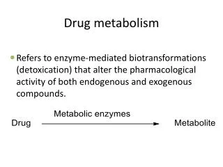
Conjugation Reactions – Some Definitions Glucuronidation “conjugation” is the addition of a molecule to the drug or xenobiotic GSH conjugation GSH = glutathione (a tripeptide derivative) Sulfate conjugation Amino acid conjugation Acetylation Methylation “Phase II metabolism” is terminology coined by RT Williams, whereby a compound is first subject to oxidation reduction or hydrolysis (Phase I reactions), which may be associated with bioactivation, and then the functional group created is conjugated to a less toxic or inactive compound. (the product is not always less toxic or inactive). Some confusing terminology, both related to drugs: Phase 1, Phase 2, Phase 3 are designations used in the sequence of drug development and evaluation by FDA in the US. Phase I and Phase II applied to describe types of drug metabolism was first used in the 1940s.
Classic example with conjugation occurring after introduction of a nucleophilic site (phenol) • 5-MeO-DIPT is a hallucinogen/stimulate that is DEA Class-I as a drug of abuse. Kamata et al., Drug Metabolism Disposition, 34:281-287, 2006
Many compounds or drugs contain functional groups that can be directly conjugated and thus do not require Phase I metabolism to create “handles” (nucleophiles) for conjugation. Ciprofloxacin Propranolol Bilirubin Diflunisal Acetaminophen Thyroxine (T4) Q. What are the nucleophilic functional groups in the drugs and endogenous compounds above?
Conjugation does not always result in less toxicity or inactivation, eg. Morphine-6-glucuronide is pharmacologically active N-acetyl procainamide is pharmacologically active GSH conjugates of haloalkenes are nephrotoxic via b-lyase Acylglucuronides are reactive, binding covalently to proteins • Of 52 pcol active metabolites in a 1985 review, only two were conjugates and those were acetylated products (Sutfin and Jusko, in Drug Metabolism and Disposition: Considerations in Clinical Pharmacology, Wilkinson and Rawlins, ed. MTP Press, Boston, 1985). • Conjugated metabolites that are pharmacologically active are not often successfully developed into drugs, whereas there are many examples of active Phase I metabolites that become drugs. (A. Fura, Drug Discovery Today, 2006).
TABLE 2 Active metabolites developed as drugs Metabolite drugs Parent drugs Acetaminophen Phenacetin Cetirizine Hydroxyzine Desimipramine Imipramine Desloratadine Loratadine Digoxin β-methyldigoxin Fexofenadine Terfenadine (withdrawn due to cardiotoxicity) Mesoridazone Thioridazone Morphine Codeine Nortriptyline Amitriptyline Oxazepam Diazepam Phenobarabital Primidone N-Acetylprocainamide Procainamide (A. Fura, Drug Discovery Today, 2006) All of these examples are Phase I metabolism with slightly more hydrophilic metabolites or are conjugation reactions that yield more lipophilic metabolites (codeine is methylmorphine; N-acetylprocainamide is an acetylated form of procainamide, thus examples of Phase II metabolism) Most Phase II metabolism yields metabolites that are polar with poor properties for bioavailability after oral administration. High polarity of the conjugate usually reduced cell wall permeability.
Mophine 6-glucuronide was discovered in 1969 to have analgesic activity, then more fully evaluated in the late 1980s and 1990s. Subsequent studies were conducted in humans for its use as a potential drug. morphine - 6 - glucuronide • Questions to consider: • Physical properties of M6G relative to M? • Would M6G be expected to have good oral bioavailability? • Would M6G likely cross the blood brain barrrier, BBB, passively? • Volume of distribution for M6G relative to M?
Common features of Phase II metabolism • Coupling of a conjugate: • Increase in molecular weight of the product. Conjugate D MW pKa range. glucuronidation 176 3 - 3.5 glycine conjugation 57 3.5 - 4.0 sulfation 81 < 1 glutathione conjugation 289 2.1, 3.5 methylation 14 neutral N-acetylation 42 neutral • Decrease (more common) orincrease in lipophilicity of the metabolic product relative to the “parent compound”. • Acidic conjugate may increase binding to albumin, thus decreasing V (eg. acetaminophen and triamterene sulfates). • Increased polarity of conjugate may limit passive partitioning into cells, thus decreasing V.
Conjugated metabolites often have higher clearance than the parent drug, due in part to active excretion into urine and/or bile. • Would a metabolite be expected to have a half-life longer or shorter than that of the parent drug? (Consider the half-life equation and the rate-limiting step when there is a sequential series of steps). • Usually there is little definitive PK of metabolites (CL, V, t1/2 ) in humans or preclinical species unless metabolite is found to be active and leads to drug candidate status. Newer guidelines require more info on metabolite toxicity and PK if the metabolite is considered a "major metabolite" by FDA guidelines. Drug (in blood) Metabolite(in blood) Metabolite in urine and bile t1/2 = ln 2 * V / CL where: V is the volume of distribution CL is the clearance of the molecule
Common features of Phase II metabolism ii. Co-substrate synthesis and availability
Common features of Phase II metabolism iii. Effect of conjugation on directing metabolite excretion. Phase II conjugation often creates anionic metabolites that are then efficiently excreted into the bile via transporters. Active secretion of organic acid metabolites in the renal proximal tubules also enhances excretion. MRP2 hepatic transporter UGT1A1 • Neonates often have immature levels of glucuronidation, thus unconjugatedhyperbilirubinemia. • Gunn rat which lacks UGT1*1 also develops unconjugatedhyperbilirubinemia. • Simple bile duct blockage usually causes increased conjugatedbilirubin.
Common features of Phase II metabolism iii. Effect of conjugation on directing metabolite excretion. There is a qualitative increase in biliary excretion of compounds with higher molecular weight (Hirom 1972, Klaassen, 1981), thus conjugation to metabolites that are more polar and ionic than the parent drug often enhances the bile/plasma ratio of a metabolite relative the parent drug. Shown here is data of drugs administered to rats where the “biliary excretion threshold” appears to be about 325 daltons. Estimates for humans, based upon fraction of drug excreted in urine and assuming mostly hepatic metabolism, suggest a higher threshold of 400-500 daltons for favorable biliary vs renal excretion of small organic molecules. (Hirom, PC, Biochem J. 129, 1071 (1972). • More recently many drugs have larger MW, thus there seems to be an increasing importance in the role of biliary excretion on drug disposition, e.g. irinotecan, MW=587
Common features of Phase II metabolism iv. Deconjugation possible , which can result in reversible metabolism in vivo. e.g. Disposition of zomepirac acyl glucuronide (ZG) when given intravenously to rats. The ester glucuronides are labile to esterases/hydrolases in vivo zomepirac (NSAID, withdrawn due to toxicity, anaphylaxis)
Common features of Phase II metabolism • Deconjugation possible, which can result in reversible metabolism in vivo. • Sulfates and acylglucuronides can hydrolyze within physiological pH range, eg. diflunisal sulfate, ketorolacacylglucuronide. • Glucuronides are susceptible to b-glucuronidase cleavage in the gi tract, thus undergoing enterohepatic recycling - reversible metabolism. b-Glucuronidase in the gut is primarily a product of enteric bacteria. • Acylglucuronides(ester functional group) are often hydrolyzed by esterases in vivo via liver and blood. • Glycine conjugates and acetylation products are subject to possible cleavage by hydrolases/ esterases in vivo.
Common features of Phase II metabolism • The major Phase II enzymes, glucuronidation and sulfation, are not so highly inducible as noted for P450s. Only several fold induction has been noted in animals or humans. • Induction increases the elimination clearance of a xenobiotic or endogenous compound, thus reducing steady state levels and makes optimization of dosing more difficult. • Induction is problematic in drug development and an undesirable characteristic for a drug, thus drugs that have Phase II metabolism as their primary route of elimination may be easier to develop.
UGTs and the Glucuronidation Reaction Smith, Phase II Metabolism, UNC Chapel Hill, 8/2012
Clearance mechanisms for the top 200 drugs prescribed in the United States in 2002 • Metabolism most important. CLr of unchanged drug is significant. Bile is more likely larger than the sliver shown due to difficulty of measuring CLbile and many newer drugs are quite high MW. • CYPs dominate, but UGTs are dominant for Phase II. Glucuronidation is also very important for sequential metabolism, after oxidation. • UGT pie is likely not so accurate and all of the active UGTs are not listed. • Establishing which UGT is dominant for a drug glucuronide formed is difficult, since tissue specific expression is not well know (certainly not in 2002). • From: Williams JA, et al. Drug Metab Dispos. 2004, 32:1201-1208 Smith, Phase II Metabolism, UNC Chapel Hill, 8/2012
UGT’s are a membrane bound enzyme located in the endoplasmic reticulum (ER) of the cell. • UGTs are in the microsomal fraction when prepared from liver and other tissues. Smith, Phase II Metabolism, UNC Chapel Hill, 8/2012
Glucuronidation is usually a detoxification pathway Though most glucuronides are inactive, they do not always result in less toxicity or inactivation, eg. morphine glucuronide is pcol active acyl glucuronides are reactive, binding covalently to proteins acetylaminofluorene hydoxylamine glucuronide is reactive • Would one expect M6G to be absorbed orally? • Q. Would M6G have the same tissue distribution as morphine? Smith, Phase II Metabolism, UNC Chapel Hill, 8/2012
Conjugation with UDPGA via the SN2 reaction requires a nucleophile or “handle” on the drug or xenobiotic Smith, Phase II Metabolism, UNC Chapel Hill, 8/2012
Stability of Glucuronide Conjugates Vary Glucuronides are excreted into bile, plasma and urine. Potential differences in glucuronide metabolites formed: i. Stability of the glucuronide products to chemical hydrolysis. UGT Q. Would the glucuronide metabolites of racemic ibuprofen be racemic? Smith, Phase II Metabolism, UNC Chapel Hill, 8/2012
Stability of Glucuronide Conjugates Vary Glucuronides are excreted into bile, plasma and urine. ii. Stability of the glucuronides to b-glucuronidase e.g, Phenylbutazone, Ethclorvinyl UGT • All glucuronides are cleaved by b-glucuronidase except C- glucuronides Smith, Phase II Metabolism, UNC Chapel Hill, 8/2012
Two major human families of UGT - UGT1 and UGT2 From: Guillemette et al., Drug Metab Rev, 2010 Smith, Phase II Metabolism, UNC Chapel Hill, 8/2012
UGT Structure and Homology Smith, Phase II Metabolism, UNC Chapel Hill, 8/2012
Biochemical pathways for cosubstrate UDP-GA • Glucose-1-P is derived from glycogen and the reactions are rapid, so co-substrate depletion not often observed unless in starved animals. • Cosubstrate depletion has been observed in animals with very large doses of drugs (e.g. acetaminophen), but recovery of UDPGA is rapid. Unlikely to occur in humans unless severe overdose occurs. • UDP-GA transport from cytosol to ER can be rate limiting in vitro. • Some compounds can deplete UDP-GA by other mechanisms, eg. diethyl ether, halothane and phenobarbital. • UGT is a bisubstrate enzyme, thus [UDP-GA] influences rate; the conc. in vivo not known at level of ER, but saturating levels of 25-40 mM use in vitro with unactivated microsomes and 2-10 mM with activation (e.g. Triton-X, Brij35, alamethicin). Smith, Phase II Metabolism, UNC Chapel Hill, 8/2012
Methods for characterizing metabolites as glucuronides Susceptibility to b-glucuronidase. Sources of b-glucuronidase, Controls (1,4-saccharolactone, Positive Controls) ii. Release of glucuronic acid (reducing sugar) by acid or b-glucuronidase. (early methods used colorimetric rxns for glucuronic acid) iii. Spectroscopy - NMR and MS. Based catalyzed acyl migration Isomeric Conjugates Johnson CH, et al. Xenobiotica, 40: 9-23, 2010 Johnson, CH, et al. Anal Chem, 79: 8720, 2007 Smith, PC et al. Drug Metab. Dispos. 13: 110-112 1985 Smith, Phase II Metabolism, UNC Chapel Hill, 8/2012
Mass Spectrometry for ID and Quantification of Glucuronides • Mass spec primarily provides M+H + , and loss of 176 (sugar) which can also occur via in source fragmentation. • No information on the site of glucuronic acid attachment (if multiple plausible sites) or isomerization (acyl migration) is provided via MS. • Site of attachment may be possible if other fragmentations of drug occur with retention of sugar. Smith, Phase II Metabolism, UNC Chapel Hill, 8/2012
Properties of Glucuronide Conjugates • Increase in molecular weight of the product (+176). • Glucuronic acid is chiral, thus products of a racemate (many older drugs are marketed as racemic mixtures, e.g. ibuprofen) are diastereomeric glucuronides, e.g. R-glucuronide, S-glucuronide. • Adding glucuronic acid (pKa 3—3.5 alters the charge on the metabolite relative to parent drug, • Adding glucuronid acid increases the polarity, thus influencing membrane transport, tissue distribution. Typically, MRP, OATP, BCRP and OAT transporters will efflux glucuronides. Smith, Phase II Metabolism, UNC Chapel Hill, 8/2012
Reversible metabolism via enterohepatic cycling Liu and Smith, Current Drug Metabolism, 2006 EHC is not irreversible elimination, thus it acts as a distribution compartment for some drugs. Interrupting EHC by bile duct drainage can reduce the AUC and alter the disposition. IV dose of valproic acid to rats circles: Bile intact rats triangles: Bile exteriorized rats Valproic acid glucuronide Smith, Phase II Metabolism, UNC Chapel Hill, 8/2012 e.g. VPA - Pollack and Brouwer, J Pharmacokin Biopharm 19: 189, 1991
Fig. 5. Representative profile of the MPA and MPAG in a human subject after a 1 gm oral dose of MMF at the baseline period w/o antibiotics (squares) and with concomitant antibiotics, metronidazole and norfloxacin (triangles). Reversible metabolism via enterohepatic cycling O.J. NadererJ. Clin. Pharmacol. 45: 219-226 (2005). • Other means to alter EHC include: • Elimination of gut bacterial flora (see MPA example) • Inhibiting b-glucuronidase in intestine • Binding metabolite excreted in bile (e.g. cholestyramine, charcoal). Smith, Phase II Metabolism, UNC Chapel Hill, 8/2012
Reversible metabolism via in vivo cleavage –deconjugation of acylglucuronides in systemic circulation Zomepirac Liu and Smith, Current Drug Metabolism, 2006 • Acyl glucuronides can be very labile once produced and exposed to hydrolases in vivo. • PMSF and other nonspecific esterases have been shown to inhibit this cleavage • The unproductive conjugation increases exposure to the parent drug in vivo. • Inhibition of CL21 should increase the apparent clearance of the drug, thus decreasing drug exposure. Smith, Phase II Metabolism, UNC Chapel Hill, 8/2012
Example of deconjugation of acylglucuronides in systemic circulation in humans Valproic acid glucuronide – an acyl glucuronide (data in monkeys) Doripenem (S-4661) • Inhibition of valproic acid glucuronide cleavage in vivo carbapenem antibiotics, thus increasing the clearance of VPA, and decreasing the exposure to VPA is observed in humans resulting in failure of antisiezure medication. • Putative inhibition of systemic cleavage of VPA-glucuronide by carbapenem blocking hydrolytic enzymes, (possibly carbamoylation of the serine active site of the enzymes?) • Kojima S, et al. Antimicrob Agents Chemotherapy ,1998. • Nakajima et al. Drug Metab Dispos, 32: 1383, 2004 Smith, Phase II Metabolism, UNC Chapel Hill, 8/2012
AcylGlucuronides are Unstable and Reactive Smith, Phase II Metabolism, UNC Chapel Hill, 8/2012
AcylGlucuronides are Unstable and Reactive Smith, Phase II Metabolism, UNC Chapel Hill, 8/2012
Are AcylGlucuronides Really Toxic? From FDA, http://www.fda.gov/OHRMS/DOCKETS/98fr/FDA-2008-D-0065-GDL.pdf • Although some acyl glucuronides are reactive in vitro and adducts are • detected in vivo, there is no conclusive link that they are the cause of toxicity. • eg. Ibuprofen forms adducts in vivo. M. Castillo, et al. Clin. Pharmacol Ther. 57: 636-644, 1995. Smith, Phase II Metabolism, UNC Chapel Hill, 8/2012
Location of UGTs in the body • Liver: Most important organ with respect to tissue levels and range of UGT’s present. • Kidney: Has UGT’s for some substrates, e.g. human kidney, but not rat, can form morphine-3-glucuronide; in rabbit, proximal tubule had highest level of UGTs. • Renal metabolism is suspected to conjugate some drugs when large amount of glucuronide are found in urine, but plasma levels are not measurable (thus CLR greatly exceeds blood flow), eg. ketorolac. • The gi tract also has UGTs, though their role in first-pass absorption is not yet fully understood. • (Tukey RH and Strassburg CP, Mol. Pharmacol. 59: 405-414 (2001)) • Many other tissues have UGTs, though isozyme distribution and activities vary. Smith, Phase II Metabolism, UNC Chapel Hill, 8/2012
Location of UGTs in the body LC-MS of UGT Proteotypic Peptides Hepatic UGTs 0 10 time (min) 20 30 • Antibodies are not available for many UGTs due to high homology, nor are the ones available always very specific. • mRNA supports diverse UGT2B isoform disposition in tissues. (Ohno S, Nakajin S, Drug MetabDispos, 37: 32-40, 2009), though mRNA and protein expression are not always well correlated, especially when comparing isoforms. • Quantitative proteomics allows the measurement of proteins via LC-MS analysis of their respective proteotypic (i.e. unique signature) peptides. • 10 UGT isoforms in human liver - UGT2B17 absent in about 30% human livers (1A and 2B families). • 4 isoforms in human intestine, UGT1A1, 1A10, 2B7, 2B17. • 3 isoforms in human kidney, UGT1A6, 1A9 and 2B7. • No UGT isoforms were detected in human lung. • Specific expression of UGTs in tissues, e.g. cancer, may be a mechanism for tissue resistance to some drugs or to modulate tissue specific exposure to endogenous compounds such as estrogens. Harbourt DE et al. Anal Chem, 2011. Fallon JK et al. Drug Metab Letters, 2: 210-222, 2008. Smith, Phase II Metabolism, UNC Chapel Hill, 8/2012
Species Differences in UGTs • Species differences are common due to differences in UGT structure and locations, e.g. rat kidney, but not human, conjugates bilirubin. • Most often cited difference is the low level of some UGTs in the cat which makes it sensitive to some planar phenol such as acetaminophen and salicylate. Defect appears to be on Exon 1 for UGT1A6, with identification of 2 stop codons and 3 deletions resulting in frame shifts, thus UGT1A6 is a pseudogene in cats (and other species?). • Court MH and Greenblatt DJ. Pharmacogenetics 10: 355-369 (2000). • Guinea pig is sometimes stated to have a high efficiency for glucuronidation. • Formation of quanternary amine glucuronides, once thought only to occur in man, has been noted in guinea pig and rabbit. • Gunn rat is lacking UGT1A’s and thus unable to conjugate bilirubin as well as many other substrates that are glucuronidated in normal rats. Smith, Phase II Metabolism, UNC Chapel Hill, 8/2012
Species Differences in UGTs Cats of all types appear to lack Ugt1A6 (pseudozyme) which may be due to cats being “requisite carnivores”, thus evolution removed an enzyme that was not needed. Thus, cats are sensitive to aspirin and acetaminophen that rely on glucuronidation for elimination. Cats would not be a good model for drug development. Another requisite carnivore that lacks Ugt1A6 is the hyena. Also the Northern Elephant Seal : Shrestha, B et al & Court, M. (2011PLoS ONE, 6 (3) DOI: 10.1371/journal.pone.0018046 Smith, Phase II Metabolism, UNC Chapel Hill, 8/2012
Inducers of Glucuronidation Induction of UGTs, and other Phase II enzymes is usually modest, several fold, in contrast to possible induction noted for P450s. General inducers: PAH analogs, such as 3-methylcholanthrene and b-naphthoflavone, are not very specific for UGT isozymes and also induce P450s. Phase II selective inducers are reported to activate the Antioxidant Response Element (ARE), e.g. BHA, oltipraz, 1,7-phenanthroline, and induce UGTs, GSH-transferases and sulfation without any apparent effect on oxidative metabolism. Lamb JG and Franklin MR. Drug Metabol. Dispos. 28: 1018-10-23 (2000). Studies have shown the involvement of xenobiotic response elements in UGTs and that UGTs are inducible by activators/ligands of PXR, CAR, PPAR and AhR . Yueh MF, e al. J Biol Chem. 278:15001-15006 (2003). Mackenzie PI, et al. Curr Drug Metab. 4:249-257 (2003). Zhou J, Zhang J, Xie W. Current Drug Metab. 6: 289-298 (2005). Buckley DB, Klaassen, Drug Metab. Dispos. 37: 847 (2009). Review of induction for UGTs, see: Remmel RP, Zhou J, Argikar UA, UDP-Glucuronosyltransferases. In: PG Pearson, L.C. Wienkers, eds., Handbook of Drug Metabolism, 2nd Ed., Informa Healthcare, New York, 2009. Smith, Phase II Metabolism, UNC Chapel Hill, 8/2012
UGT Polymorphism To date, deficient conjugation with bilirubin which leads to severe disease (Crigler Najar) or elevated bilirubin (Gilberts syndrome) have been identified. • Crigler-Najar is commonly associated with a 13 bp deletion in exon 2 of UGT1*1. • Gilberts syndrome is commonly associated with a defect in the promotor region of exon 1. • Normals: A(TAT)6TAA; Gilbert’s: A(TAT)XTAA, where X=7, 8. Smith, Phase II Metabolism, UNC Chapel Hill, 8/2012
Gilberts Syndrome : Severe Neutropenia Risk: 7/7 vs 6/6 + 6/7 Genotypes Unadjusted Odds Ratio From Parodi et al, FDA Subcommittee presentation, November, 2004 Product label for Camptostar (irinotecan): “Individuals who are homozygous for the UGT1A1*28 allele are at increased risk for neutropenia following initiation of CAMPTOSAR treatment. A reduced initial dose should be considered for patients known to be homozygous for the UGT1A1*28 allele” “However, the precise dose reduction in this patient population is not known” Smith, Phase II Metabolism, UNC Chapel Hill, 8/2012
Other polymorphisms of UGT due to random genetic differences have been reported, but only a few have suggested to influence drug metabolism in the clinic. e.g. variants of UGT1A9 that appear to create a fast metabolizer phenotype in 15% patients. Polymorphisms in UGT1A9 promoter (T-275A and C-2152T) associated with increased hepatic 1A9 protein expression (Girard et al. 2004) have shown to decrease AUC of mycophenolic acid (immunosuppressive) when patient had either or both polymorphisms (Kuypers et al., 2005) Non-carriers of the T-275A and C-2152T 1A9 promoter polymorphisms (low 1A9 expressors) showed a trend toward increased gastrointestinal toxicity of mycophenolic acid. For a comprehensive listing of SNPs for UGTs, see: http://www.flinders.edu.au/medicine/sites/clinical-pharmacology/ugt-homepage.cfm Smith, Phase II Metabolism, UNC Chapel Hill, 8/2012
= ATP Sulfurylase, = APS Kinase Bifunctional Enzyme in Mammals – PAPS Synthetase (2 isoforms) M.Coughtrie, U. Dundee Sulfotransferases (SULTs) Co-factor is PAPS (3’-phosphoadenosine 5’-phosphosulfate) – a high energy intermediate. Low levels in vivo, but synthesized quickly from inorganic sulfate or catabolism of cysteine and methionine. Cosubstrate Depletion. Reduced inorganic sulfate or cysteine (secondary to GSH depletion) can cause co-substrate-dependent decrease in sulfation rates. e.g. high doses of acetaminophen or harmol (Levy and Morris, Pang) Smith, Phase II Metabolism, UNC Chapel Hill, 8/2012
Sulfotransferases (SULTs) Location: High levels in the liver, notable in intestine, common throughout the body. A cytosolic enzyme, not membrane bound. Induction: Little evidence for induction of sulfotransferases in vivo with classic inducers Pb, BHA or 3MC, but PCN does induce (Liu, Klaassen Drug Metab Dispos. 24: 85 (1996). Induction by PCN is regulated by PXR (Sonoda, J, R. Evans et al., PNAS 99: 13801-6, (2002)). M. Coughtrie, U. Dundee Smith, Phase II Metabolism, UNC Chapel Hill, 8/2012
Substrates for Sulfotransferases (SULTs) minoxidil • Phenols and analine type amines are sulfated • Frequently phenols have both glucuronidation • and sulfation as competing conjugation rxn. • Sulfation is often a high affinity, low capacity • pathway, while glucuronidation is frequently • low affinity, high capacity. • Thus, at low dose, sulfation may dominate, • but as dose increases, glucuronidation can • become the major route. • Sulfates are frequently excreted in urine, though • some bile acid sulfates are excreted into bile. Smith, Phase II Metabolism, UNC Chapel Hill, 8/2012
Properties of Sulfate Metabolites • Usually inactive metabolites, but minoxidil sulfate is active. • Sulfates are strong acids (pKa < 1) • As an acid, sulfates often bind to albumin. Plasma protein binding of sulfate metabolite • can be higher than that of the parent. • e.g. acetaminophen fb =0% (in sheep; <20% in humans) • “ sulfate fb = 36% • “ glucuronide fb = 4% • 4-methyl umbelliferone fb =90% • “ sulfate fb = 97% • Sulfate metabolites are usually excreted by the kidney, though for larger molecules, • sulfates can be excreted in the bile. • Sulfation is subject to possible reversible metabolism, eg. Diflunisal, which is a pH- • dependent process (more labile at lower pH). • Sulfation of hydroxylated aromatic amines (e.g. acetylaminofluorene) can lead to reactive • intermediates and putative toxicity. (Banoglu E, Current Drug Metab. 1: 1-30 (2000). Smith, Phase II Metabolism, UNC Chapel Hill, 8/2012
Summary of Phase II metabolism: • Phase II conjugation usually results in a large polar functional group to be added, thus decreasing lipophilicity. • Acetylation and methylation, however, increase lipophilicity. • A nucleophilic site is needed for conjugation, some added by Phase I metabolism. • Phase II metabolites often have high clearances and lower volume of distribution relative to the “parent” compound. • Phase II metabolites are often excreted in urine and bile, sometimes at high concentrations. • Reversible metabolism is fairly common with Phase II metabolism. • Co-substrate depletion is possible, thus slowing rates of metabolism, especially in toxicology studies conducted at very high doses. • The major organs of elimination for Phase II metabolism are usually liver, kidney and intestine, though there may be important tissue specific metabolism. • The Phase II enzymes are families, often with overlapping substrate affinity, though there are some examples of very specific substrates. • Phase II metabolism is not very inducible. Smith, Glucuronidation, UNC Chapel Hill, 9/2011